The microscope : and its application to vegetable anatomy and physiology / by Dr. Hermann Schacht ; edited by Frederick Currey, M. A.
- Hermann Schacht
- Date:
- 1855
Licence: Public Domain Mark
Credit: The microscope : and its application to vegetable anatomy and physiology / by Dr. Hermann Schacht ; edited by Frederick Currey, M. A. Source: Wellcome Collection.
194/230 (page 170)
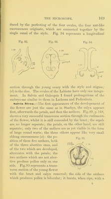
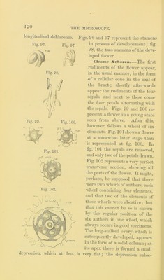
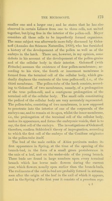
![longitudinal dehiscence. Fig. 96. Fig. 97. depression, which at first is Figs. 9G and 97 represent the stamens in j:)rocess of develo2)ement; fig. 98, the two stamens of the deve- lo2>ed flower. Ai-l>orea. The first rudiments of the flower ai)j>ear, ui the usual manner, in the form of a cellular cone in the axil of the hi’act; shortly afterwards a])jiear the rudiments of the four se])als, and next to these come the four petals alternating Avith the sepals. Figs. 99 and 100 re- present a flower in a yoiing state seen from above. After this, however, follows a whorl of six elements. Fig. lOlshoAvsaflower at a somewhat later stage than is represented at fig. 100. In fig. 101 the sepals are removed, and only two of the petals drawn. Fig. 102 represents a very perfect ti’ansA^erse section, shoAving all the jAai-ts of the floAvei*. It might, perhaps, be sujiposed that there Avere tAvo Avhorls of anthers, each whorl containing four elements, and that tAvo of the elements of these AA'horls AA'ere aljortiA'e; but that this cannot be so is shoAAui by the regular jAosition of the six anthei-s in one Avhorl, Avhich ahvays occurs in good specimens. The long-stalked o\-ary, Avhich is subsequently deA’elojied, ajipeai’s in the form of a solid cohmui; at its apex there is formed a small very flat; the depi-cssion subse-](https://iiif.wellcomecollection.org/image/b28071761_0194.jp2/full/800%2C/0/default.jpg)