Cancer of the uterus : its pathology, symptomatology, diagnosis, and treatment : also the pathology of diseases of the endometrium / by Thomas Stephen Cullen ; with eleven lithographic plates and over three hundred coloured and black illustrations in the text by May Brödel and Hermann Becker.
- Thomas Stephen Cullen
- Date:
- 1900
Licence: Public Domain Mark
Credit: Cancer of the uterus : its pathology, symptomatology, diagnosis, and treatment : also the pathology of diseases of the endometrium / by Thomas Stephen Cullen ; with eleven lithographic plates and over three hundred coloured and black illustrations in the text by May Brödel and Hermann Becker. Source: Wellcome Collection.
Provider: This material has been provided by the Francis A. Countway Library of Medicine, through the Medical Heritage Library. The original may be consulted at the Francis A. Countway Library of Medicine, Harvard Medical School.
55/774 (page 33)
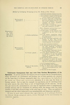
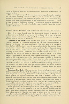
![Maeroscopical appearances. Examination of scrap- | ings from J the body of I the uterus. A. Surface epithelium. Method of studying Scrapings from the Body of the Uterus (' a. Estimate quantity of tissue removed. I b. How large are the individual pieces ? | c. Is it possible to make out the smooth velvety surface of the mucosa ? d. Are there finger-like or branching particles springing from the sur- face of the scraping 1 If so, note carefully the blood capillaries ramifying over them. e. Is the tissue of brain-like appear- ance and consistence, or not? Note.-—Examine sections from vari- ous pieces of scrapings. 1. Is the surface of the mucosa even, or gathered up into folds or pa- pillae ? 2. Is the surface epithelium intact and one layer in thickness, or has it proliferated ? 3. Are the individual epithelial cells normal, or have they become swollen ? Do they contain an in- creased amount of chromatin ? 1. Are they normal in number? 2. Do they present the usual shape, or are they markedly convoluted and bunched together! Is there much branching? 3. Are the glands uniformly distrib- uted throughout the stroma? B. Uterine glands... \ 4. Is the gland epithelium one layer in thickness, or are the glands partly or completely filled by epithelial cells? 5. Describe the gland contents. 6. If perchance a piece of muscle should be attached to the scrap- ing, does it show gland invasion ? f 1. Is it of normal density? | 2. Are the stroma cells swollen? J 3. Does it show much small-round- ] cell or polymorphonuclear infil- tration ? Describe its blood supply. Microscopical examination. Stroma of the uterine mucosa. 14. Unfortunate Consequences that may arise from Careless Manipulation of the Specimens.—In private practice the dangers are not so great, but in a hospital,where many operations are performed, and perhaps two or three patients are curetted on the same day, it is always imperative that the specimens be labelled as soon as they are received, otherwise it is very easy for them to become mixed. Let us take for example two specimens, one of curettings from an early cancer, the other from a case of hypertrophy of the mucosa. If the two get mixed, the carcinomatous case may be reported to have a comparatively normal mucosa, and no treatment be advised, while the woman with but little trouble is told that a hysterectomy is the only hope of cure. Thus one patient is doomed, and the other subjected to an unwarrantable and dangerous operation. If one is not sure about the specimens, it is always better to throw them away at once, even though it be necessary to curette again. 4](https://iiif.wellcomecollection.org/image/b21047984_0055.jp2/full/800%2C/0/default.jpg)