Licence: Public Domain Mark
Credit: On inflammation / by G. Thin. Source: Wellcome Collection.
Provider: This material has been provided by The Royal College of Surgeons of England. The original may be consulted at The Royal College of Surgeons of England.
25/88 page 23
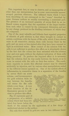
![cornea injured by the caustic, and differing from tlie other parts by the abundant deposit of the gold chloride, teaches us two im- portant facts in regard to inflammation, namely, that inflammatory change produced by injury at a given point may be propagated from that ])oint along an isolated well-demarcated tract towards the nearest bloodvessels ; and that further, in an early stage of inflam- mation, the nature of the fluid present in an inflamed tract differs chemically from that present in the surrounding uninflamed tissue. The strength of this inference lies in the weight assigned to the reduction in the tissues of the chloride of gold in the form of a dark purple precipitate. I am unable to state what the substance is, the presence of which is necessary to the chemical action indicated by the precipitate. We know that it is unequally distributed in the tissues. It is notably present in ganglion cells, nerve fibres, and in tlie protoplasm which enters into the composition of stellate cells in some forms and stages of connective tissue. It exists nor- mally to a certain extent in the lymph fluid of the tissues, and is always present in that of the cornea to a greater or less degree. But when it is present in an inflamed tract in larger quantity than in the uninflamed portions of the same cornea, it becomes certain that the lymph fluid in an inflamed tissue differs chemically from that usually present in the same tissue in a normal condition. Observations by pathologists, made by other methods in other tissues, indicate that the change consists in the presence in the fluid of a fibrinogenic material, and the direction of the tract towards the conjunctiva indicates the nearest bloodvessel as its source. Fig. 20 represents, as magnified by a low power, a preparation obtained by cauterization of the cornea of a winter frog {Rana esc), which was killed twelve hours after- wards, and the cornea treated by gold. In the figure the dark diffused appearance result- Fio. 20.-InfIame<l tract in the comca of a winter frog ing frOm the Cauterization (/fi/ia ««<;.), twelve hours after cauterization. Gold prepnra- . tion. a, Bordi-r of tract, b, Cauterized surface, c, Nerve IS shoWU at b One of trunk.—Magii. 50 diam. ■ --^ ^ the borders of the inflamed tract is seen at a. The dark masses in the tract are the spaces filled by the gold precipitate. The rest of the cornea was scarcely stained at all, showing only the ramifications of the larger corneal nerves, one of which is indicated at c.](https://iiif.wellcomecollection.org/image/b22292743_0027.jp2/full/800%2C/0/default.jpg)

 No text description is available for this image
 No text description is available for this image
 No text description is available for this image