Licence: Public Domain Mark
Credit: On inflammation / by G. Thin. Source: Wellcome Collection.
Provider: This material has been provided by The Royal College of Surgeons of England. The original may be consulted at The Royal College of Surgeons of England.
49/88 page 47
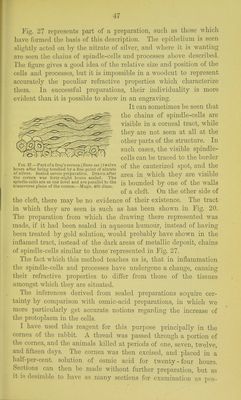
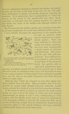
![Fig. 27 represents part of a preparation, sucli as those which have formed tlie basis of this description. Tlie e]iitlielium is seen sliglitly acted on by the nitrate of silver, and where it is wanting are seen the chains of spindle-cells and processes above described. The fignre gives a good idea of the relative size and position of the cells and processes, but it is impossible in a woodcut to represent accurately the peculiar refractive properties which characterize them. In successful preparations, their individuality is more evident than it is possible to show in an engraving. It can sometimes be seen that tlie chains of spindle-cells are visible in a corneal tract, while they are not seen at all at the other parts of the structure. In such cases, the visible spindle- cells can be traced to the border !• 10.27.-Partofa frog's cornea (;?anaejO twelve j^]^Q cautCrized SpOt, and the hours after being touched by a fine point of nitrate ^ ' of silver. Sealed scriini preparation. Drawn after gj.Q^ wMch theV are visible tlie cornea was forty-eight hours sealed. The - spindle-cells are at one level and are parallel to the ig hounded bv OUe of the Walls transverse plane of the cornea.—Magn. 400 diam. of a cleft. On the other side of the cleft, there may be no evidence of their existence. The tract in which they are seen is such as has been shoAvn in Fig. 20. The preparation from which the drawing there represented was made, if it had been sealed in aqueous humour, instead of having been treated by gold solution, would probably have shown in the inflamed tract, instead of the dark areas of metallic deposit, chains of spindle-cells similar to those represented in Fig. 27. The fact which this method teaches us is, that in inflammation the spindle-cells and processes have undergone a change, causing their refractive properties to differ from those of the tissues amongst which they are situated. The inferences derived from sealed preparations acquire cer- tainty by comparison with osmic-acid preparations, in which we more particularly get accurate notions regarding the increase of the protoplasm in the cells. I have used this reagent for tliis purpose principally in the cornea of the rabbit. A thread was passed through a portion of the cornea, and the animals killed at periods of one, seven, twelve, and fifteen days. The cornea was then excised, and placed in a half-per-cent. solution of osmic acid for twenty-four hours. Sections can then be made without furtlier preparation, but as it IS desirable to liavo as inany sections for oxnminatimi ns ]in,s-](https://iiif.wellcomecollection.org/image/b22292743_0051.jp2/full/800%2C/0/default.jpg)
 No text description is available for this image
 No text description is available for this image
 No text description is available for this image