Licence: Public Domain Mark
Credit: On inflammation / by G. Thin. Source: Wellcome Collection.
Provider: This material has been provided by The Royal College of Surgeons of England. The original may be consulted at The Royal College of Surgeons of England.
84/88 (page 82)
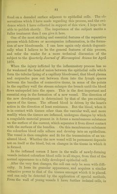
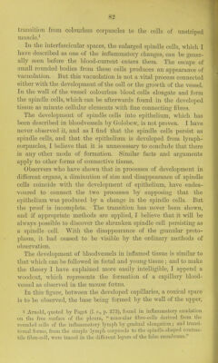
![transition from colourless corixiscles to the cells of unstriped nuiscle/ In the interfascicular spaces, the enlarged spindle cells, which I have described as one of the inflammatory changes, can be gener- ally seen before the blood-current enters them. The escape of small rounded bodies from these cells produces an appearance of vacuolation. But this vacuolation is not a vital process connected either with the development of the cell or the growth of the vessel. In the wall of the vessel colourless blood cells elongate and form the spindle cells, which can be afterwards found in the developed tissue as minute cellular elements with fine connecting fibres. The development of spindle cells into epithelium, which has been described in bloodvessels by Golobew, is not proven. I have never observed it, and as I find that the spindle cells persist as spindle cells, and that the epithelium is developed from lymph- corpuscles, I believe that it is unnecessary to conclude that there is any other mode of formation. Similar facts and arguments apply to other forms of connective tissue. Observers who have shown that in processes of development in different organs, a diminution of size and disappearance of spindle cells coincide with the development of epithelium, have endea- voured to connect the two processes by supposing that the epithelium was produced by a change in the spindle cells. But the proof is incomplete. The transition has never been shown, and if appropriate methods are applied, I believe that it will be always possible to discover the shrunken spindle cell persisting as a spindle cell. With the disappearance of the granular proto- plasm, it had ceased to be visible by the ordinary methods of observation. The development of bloodvessels in inflamed tissue is similar to that which can be followed in foetal and young tissue ; and to make the theory I have explained more easily intelligible, I append a woodcut, Avhich represents the formation of a capillary blood- vessel as observed in the mouse foetus. In this figure, between the developed capillaries, a conical space is to be observed, the base being formed by the Avail of the upper, 1 Arnold, (luoted by Paget {I. c, p. 273), found in inflammatoiy exudation on the free surface of the pleura, muscular fibre-cells derived from the rounded cells of the inflammatory lymph by gradual elongation; and transi- tional forms, from the simple Ijanph corpuscle to the spindle-sha]3ed contrac- tile fibre-cell, were traced in tlic Lliflerunt layers of the false membrane.](https://iiif.wellcomecollection.org/image/b22292743_0086.jp2/full/800%2C/0/default.jpg)