Clinical diagnosis : the bacteriological, chemical, and microscopical evidence of disease / by Rudolf v. Jaksch ; translated from the second German edition by James Cagney ; with an appendix by Wm. Stirling.
- Cagney James.
- Date:
- 1890
Licence: Public Domain Mark
Credit: Clinical diagnosis : the bacteriological, chemical, and microscopical evidence of disease / by Rudolf v. Jaksch ; translated from the second German edition by James Cagney ; with an appendix by Wm. Stirling. Source: Wellcome Collection.
Provider: This material has been provided by the Royal College of Physicians of Edinburgh. The original may be consulted at the Royal College of Physicians of Edinburgh.
71/432 (page 43)
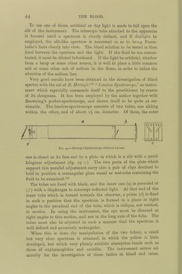
![animals, notabl}' dogs. The opinion of the latter observers finds support in a clinical notice of Lenhcniz, and also in a pathological observation recorded by II. Ilammei'.^-^ Chlorate of potash may be easily detected by spectrum analysis in fairly dilute solutions of htcmoglobin, and the spectrum of metha;- moglobin, if present, will afford presumptive evidence of the poison. Methsemoglobin is produced also by the inhalation of nitrite of amyl [Gamgee) and the injection of sodium nitrite into the blood-vessels {Hoppe-Seyle)'), as well as by kairin, thallin, hydrochiuon, pyro- catechin, iodine, bromine, turpentine, mther, perosmic acid, perman- ganate of potash {G. Ilayem) and antifebrin {Fr. [The nitrites form a compound with its oxygen, more firmly fixed than that of the oxygen in oxyhsemoglobin. They consequently tend to stop internal respiration, but are more readily displaced by the products of asphyxia than is carbonic oxide hEemoglobin, and so again permit the aeration of the blood at the lungs.] 6. Poisoning with Nitrobenzol.—It has been asserted that in dogs poisoned. with nitrobenzol the spectroscope has shown blood-changes attributable to the presence of hsematin. It would seem, then, that in any case of suspected poisoning by this means in the human subject the blood should be examined in this direction by the spectroscope. 7. Haemoglobinaemia.123—]3y this term is meant the condition in which haemoglobin is found dissolved in the blood. It is followed by Haemoglohinuria, whenever the spleen and the liver are unequal to the task of eliminating the materials derived from the destruction of the red blood-corpuscles within the vessels. The presence of free colouring matter in the blood may easily be determined thus:—A little of the blood, drawn from the patient by means of a cupping-glass, is placed immediately in a refrigerator, and allowed to remain there for twenty-four hours. If the blood is normal, perfectly clear yellowish-coloured serum will settle; whereas, if hsemo- globinsemia be established, there will be seen over the blood-clot a beautiful transparent ruby-red stratum. The spectroscope shows in the case of normal serum a feeble absorption-band in the blue part of the spectrum (at F), said to be due to lutein (Thudichurn); whilst with serum containing colouring matter it shows the two absorption- bands of oxyhsemoglobin. 8. Recognition of Changes in the Colouring-Matter of the Blood.— The changes in the character of the blood referred to above are chiefly to be estimated by means of the spectroscope. Very perfect little instruments for clinical u.se have been invented by Desaga of Heidel- berg, Zeiss of Jena, and Hoffmann of Paris. Browning’s spectroscope is also very suitable for the piu’pose.](https://iiif.wellcomecollection.org/image/b21699574_0071.jp2/full/800%2C/0/default.jpg)