Clinical diagnosis : the bacteriological, chemical, and microscopical evidence of disease / by Rudolf v. Jaksch ; translated from the second German edition by James Cagney ; with an appendix by Wm. Stirling.
- Cagney James.
- Date:
- 1890
Licence: Public Domain Mark
Credit: Clinical diagnosis : the bacteriological, chemical, and microscopical evidence of disease / by Rudolf v. Jaksch ; translated from the second German edition by James Cagney ; with an appendix by Wm. Stirling. Source: Wellcome Collection.
Provider: This material has been provided by the Royal College of Physicians of Edinburgh. The original may be consulted at the Royal College of Physicians of Edinburgh.
86/432 (page 58)
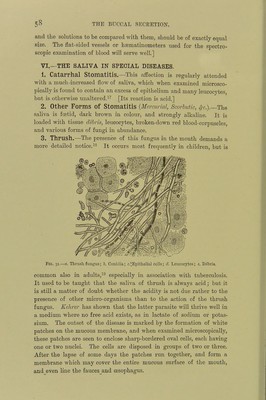
![and the solutions to be compared with them, should be of exactly equal size. The flat-sided vessels or haematinometers used for the spectro- scopic examination of blood will serve well.] VI.—THE SALIVA IN SPECIAL DISEASES. 1. Catarrhal Stomatitis.—This affection is regularly attended with a much-increased flow of saliva, which when examined microsco- pically is found to contain an excess of epithelium and many leucocytes, but is otherwise unaltered.[Its reaction is acid.] 2. Other Forms of Stomatitis {Mercurial, Scorbutic, ^-c.).-The saliva is foetid, dark brown in colour, and strongly alkaline. It is loaded with tissue debris, leucocytes, broken-down red blood-corpuscles, and various forms of fungi in abundance. 3. Thrush.—The presence of this fungus in the mouth demands a more detailed notice.occurs most frequently in children, but is Fig. 31.—a. Thrush fungus; J. Conidia; c.‘Epithelial colls; d Leucocytes; e. Debris. common also in adults,^® especially in association with tuberculosis. It used to be taught that the saliva of thrush is always acid; but it is still a matter of doubt whether the acidity is not due rather to the presence of other micro-organisms than to the action of the thrush fungus. Kehrer has shown that the latter parasite wiU thrive well in a medium where no free acid exists, as in lactate of sodium or potas- sium. The outset of the disease is marked by the formation of white patches on the mucous membrane, and when examined microscopically, these patches are seen to enclose sharp-bordered oval cells, each having one or two nuclei. The cells are disposed in groups of two or three. After the lapse of some days the patches run together, and form a membrane which may cover the entire mucous surface of the mouth, and. even line the fauces_and oesophagus.](https://iiif.wellcomecollection.org/image/b21699574_0086.jp2/full/800%2C/0/default.jpg)