Clinical diagnosis : the bacteriological, chemical, and microscopical evidence of disease / by Rudolf v. Jaksch ; translated from the second German edition by James Cagney ; with an appendix by Wm. Stirling.
- Cagney James.
- Date:
- 1890
Licence: Public Domain Mark
Credit: Clinical diagnosis : the bacteriological, chemical, and microscopical evidence of disease / by Rudolf v. Jaksch ; translated from the second German edition by James Cagney ; with an appendix by Wm. Stirling. Source: Wellcome Collection.
Provider: This material has been provided by the Royal College of Physicians of Edinburgh. The original may be consulted at the Royal College of Physicians of Edinburgh.
96/432 (page 68)
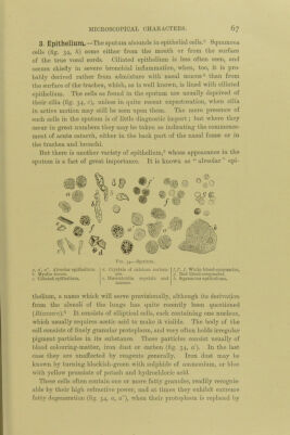
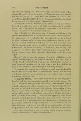
![oily drops of varying size. Sometimes large bodies like drops of fat, presumably derived from the rupture of these cells, are to be seen in the sputum (fig. 34, b). They were first observed by Virchoto,’^ who named them myelin droplets, from the resemblance they bear to similar forms produced in the destruction of nerve tissue. According to Panizza,^ however, myelin (which is only the outward form of a considerable number of different substances) is probably mucin; and in his opinion no diagnostic importance is to be attached either to myelin or to myelin-containing cells. Buhl ® thought that the appearance of alveolar epithelium in the sputum was characteristic of the disease which he has named desquama- tive pneumonia. It is certainly a fact that such cells are to be found in great profusion only in quite fresh specimens of caseous infiltra- tion of the lung, whether due to bacilli or not. But then they occur also in pneumonia, in chronic bronchitis, and in chronic pulmonary tuberculosis {Guttmann and Smidt); sometimes, too, in very large numbers. It follows from their manifestation in processes differ- ing so entirely, that their diagnostic significance is on the whole slight. \Troup believes that the presence of alveolar epithelium in the sputum belongs especially to obstinate catarrhs of the apex, when it will be found associated with columnar and ciliated cells; he observes that since such catarrhs tend in most cases to run into phthisis, we have in this a valuable and early sign of impending danger.] To examine the sputum for epithelium, a small quantity should be treated with acetic acid, when the characteristic nucleus and nucleolus will become evident; or a specimen may be stained with a watery solution of methylene blue. 4. Elastic Fibres.—These fibres occur in the sputum singly or in bundles, and they are commonly arranged in an alveolar manner (fig. 35). They are of varying length and breadth, dark-bordered, slightly curved, and generally exhibit a double contour.* Their diagnostic value is great, as a sign of serious mischief, pointing to destruction of lung tissue. They occur, accordingly, in tuberculosis, bronchiectasis, pul- monary abscess, and occasionally in pneumonia, while the other symp- toms of abscess are wanting. The author has repeatedly found elastic fibres in cases of pneumonia which otherwise ran a normal course, and he supposes that in such cases there was destruction of the pulmonary parenchyma only over a very limited area. It is a notable fact that these fibres are rarely to be met with in the expectoration of pulmo- nary gangrene, and the reason probably is that they are destroyed * The figure is taken from a case of pulmonary abscess, which was for several months under treatment in Professor Nothnagel’s clinic.](https://iiif.wellcomecollection.org/image/b21699574_0096.jp2/full/800%2C/0/default.jpg)