The mechanisms of motor end-plate depolarization due to a cholinesterase-inhibiting drug / by W.W. Douglas and W.D.M. Paton.
- Douglas, W. W.
- Date:
- [1954?]
Licence: In copyright
Credit: The mechanisms of motor end-plate depolarization due to a cholinesterase-inhibiting drug / by W.W. Douglas and W.D.M. Paton. Source: Wellcome Collection.
1/20
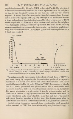
![[Reprinted from the Journal of Physiology, 1954, Vol. 124, No. 2, p. 325.] PRINTED IN GREAT BRITAIN J. Physiol. (1954) 124, 325-344 THE MECHANISMS OF MOTOR END-PLATE DEPOLARIZATION DUE TO A CHOLINESTERASE-INHIBITING DRUG By W. W. DOUGLAS and W. D. M. PATON From the National Institute for Medical Research, Mill Hill, London, N.W.I (.Received 12 November 1953) Many cholinesterase-inhibitors are stated to have a direct acetylcholine-like action as well as an action due to the preservation of acetylcholine normally destroyed. The widespread use of such drugs in analysing the functions of acetylcholine in the body depends for its validity on knowledge about such direct action. For such an action might be attributed erroneously to acetyl¬ choline; and criticisms have been levelled at analyses involving the use of anticholinesterases for this reason. In this paper we have chosen TEPP (tetraethylpyrophosphate) for study, because it is one of the strongest and most commonly used alkyl-phosphate cholinesterase inhibitors, and we have analysed its effects at the neuro¬ muscular junction. A preliminary account of this work has already been published (Douglas & Paton, 1951). METHODS I Cats anaesthetized with chloralose (80 mg/kg intravenously after induction with ethyl chloride and ether) were used in all experiments. The trachea was cannulated. Intravenous injections were made through a cannula in the jugular vein. For experiments on the depolarization of the motor end-plate, the method of Burns & Paton (1951) was used. The cat’s gracilis was exposed and enclosed in a paraffin bath formed from skin flaps, and its motor nerve tied and cut. The spatial distribution of electrical potential along a ? length of the muscle which contained suitable end-plate regions was recorded as follows: a scanning electrode was made to traverse the region chosen 15 times a minute and an indifferent electrode was placed on the tendon. The potential difference between the electrodes was applied to the Y-plates, and the movement of the scanning electrode (after conversion to a potential change) to the X-plates, of a cathode-ray oscilloscope. Supramaximal shocks of 0-5 msec duration were applied to the tied motor nerve through shielded electrodes. For experiments on the tibialis anterior, the contractions of this muscle prepared for close arterial injections were recorded with a flat spring myograph on a smoked drum. Samples of blood (3 ml.) for assay were withdrawn from a steel cannula tied into the carotid artery; or through a glass cannula inserted into the splenic vein, from which portal blood could be ■ I ft-](https://iiif.wellcomecollection.org/image/b30633928_0001.jp2/full/800%2C/0/default.jpg)