Microscopical morphology of the animal body in health and disease / by C. Heitzmann. With 380 original engravings.
- Carl Heitzmann
- Date:
- 1883
Licence: Public Domain Mark
Credit: Microscopical morphology of the animal body in health and disease / by C. Heitzmann. With 380 original engravings. Source: Wellcome Collection.
Provider: This material has been provided by the Augustus C. Long Health Sciences Library at Columbia University and Columbia University Libraries/Information Services, through the Medical Heritage Library. The original may be consulted at the the Augustus C. Long Health Sciences Library at Columbia University and Columbia University.
125/884 page 99
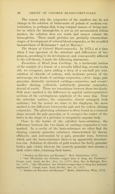
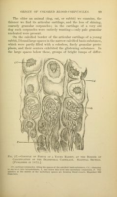
![(HU(iis OF coLoh'hn ni.()()i)-(()i{i'rscij':s. \)\) The olilor an iuiiiual (do?;, cat, oi* ral»l)it) we cxaiiiiiH', the Ihiinicf we find its articnlar <*artila}^(', and Mjc U'ss of shining, coarsely <;ranulai- coi-puseles; iu the cartilage of a very old dog such corpuscles were cntii'i^ly wanting—only ])ale gi-annlar nucleated were j)resent. On the calcified ))()rder of the articular cartilages of a young rabbit, I found large spaces in the narrow calcified l)asis-substance, which were partly filled with a colorless, finely granular proto- plasm, and their centers exhibited the glistening su1)stance. In the large spaces below these, groups of bright lumps of difter- Fir;. L'7.—Condyle of Femur of a Young Rabbit, at the Border of Calcificatiox of the Diaphyseal Cartilage. Sagittal Section. [Published in 1872.] CC, t-aitilajje-corpuscles, filling the spaces of the calcified basis-substance, CA ; changing in one level into liiiniatoblasts, X, and below this level into medullary corpuscles, P. The spindles in the middle of the medullary spaces are forniinir blood-vessels. Magnified 800 diameters.](https://iiif.wellcomecollection.org/image/b21219163_0125.jp2/full/800%2C/0/default.jpg)