Microscopical morphology of the animal body in health and disease / by C. Heitzmann. With 380 original engravings.
- Carl Heitzmann
- Date:
- 1883
Licence: Public Domain Mark
Credit: Microscopical morphology of the animal body in health and disease / by C. Heitzmann. With 380 original engravings. Source: Wellcome Collection.
Provider: This material has been provided by the Augustus C. Long Health Sciences Library at Columbia University and Columbia University Libraries/Information Services, through the Medical Heritage Library. The original may be consulted at the the Augustus C. Long Health Sciences Library at Columbia University and Columbia University.
147/884 page 121
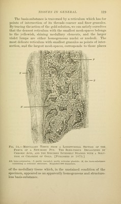
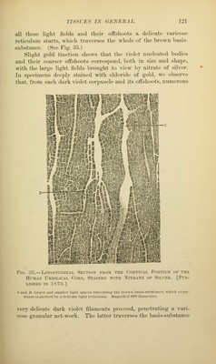
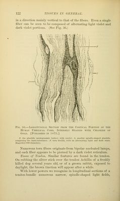
![all those liijlit fields mikI their offshoots a delicate varicose reticulum starts, wiiich traverses the whole of the lirown basis- substance. (See Fig. 35.) Sli^rht (Told tinetion shows that the violet nucleated l>odies and their coarser otlslioots correspond, both in size and shape, with the lar<;e lif^ht tields lu-ou^ht to view l)y nitrate of silver. In specinu'us deeply st<iined with chloride of gold, we observe that, from each dark v-iolet corpuscle and its offshoots, numerous Fig. 35.—Longitudinal Section from the Cortical Portion of the Human Umbilical Cord, Stained with Nitrate of Silver. [Pub- lished IN 1873.] s anil B. largtr and smaller light siiates traversing the biown basis-suhstante, whirli every- where is pierced by a delieate light retictUinn. Miagnitteil 800 diameters. very delicate dark violet filaments proseed, penetrating a vari- cose gi-anular net-work. The latter traverses the basis-substance](https://iiif.wellcomecollection.org/image/b21219163_0147.jp2/full/800%2C/0/default.jpg)
 No text description is available for this image
 No text description is available for this image
 No text description is available for this image