Microscopical morphology of the animal body in health and disease / by C. Heitzmann. With 380 original engravings.
- Carl Heitzmann
- Date:
- 1883
Licence: Public Domain Mark
Credit: Microscopical morphology of the animal body in health and disease / by C. Heitzmann. With 380 original engravings. Source: Wellcome Collection.
Provider: This material has been provided by the Augustus C. Long Health Sciences Library at Columbia University and Columbia University Libraries/Information Services, through the Medical Heritage Library. The original may be consulted at the the Augustus C. Long Health Sciences Library at Columbia University and Columbia University.
148/884 page 122
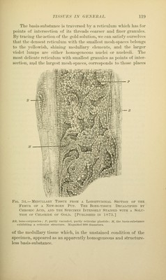
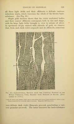
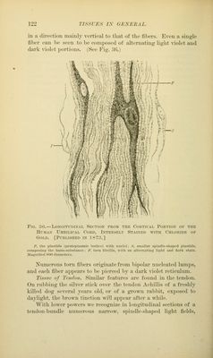
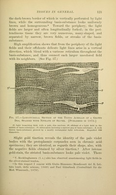
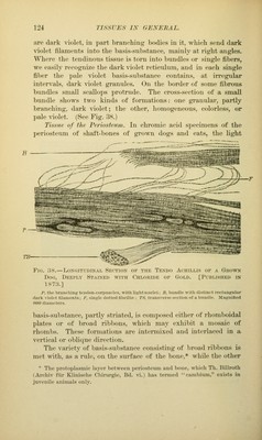
![in a dii-ectiou mainly vertical to that of the fibers. Even a single fiber can be seen to be composed of alternating light \-iolet and dark violet portions. (See Fig. 36.) Fig. 36.—LoxGiTrniXAL Section from the Cortical Portion of the Human Umbilical Cord, Intensely Stained with Chloride of Gold. [Published in 1873.] P, the i)lastiils (protoplasmic bodies) with nuclei; S, siualler spindle-shaped plastids, connposinj; the liasis-suhstance; F, torn tibrilla, with an alternating light and dark stain. MagniJied 800 diameters. Numerous torn fibers originate fi-om bipolar nucleated lumps, and each fiber appears to be pierced by a dark violet reticulum. Tissue of Tendon. Similar features are found in the tendon. On rubbing the silver stick over the tendon Achillis of a freshly killed dog several years old, or of a gi'own rabbit, exposed to daylight, the brown tinction will appear after a while. With lower powers we recognize in longitudinal sections of a tendon-bundle numerous narrow, spindle-shaped light fields,](https://iiif.wellcomecollection.org/image/b21219163_0148.jp2/full/800%2C/0/default.jpg)
 No text description is available for this image
 No text description is available for this image
 No text description is available for this image