Microscopical morphology of the animal body in health and disease / by C. Heitzmann. With 380 original engravings.
- Carl Heitzmann
- Date:
- 1883
Licence: Public Domain Mark
Credit: Microscopical morphology of the animal body in health and disease / by C. Heitzmann. With 380 original engravings. Source: Wellcome Collection.
Provider: This material has been provided by the Augustus C. Long Health Sciences Library at Columbia University and Columbia University Libraries/Information Services, through the Medical Heritage Library. The original may be consulted at the the Augustus C. Long Health Sciences Library at Columbia University and Columbia University.
49/884 page 23
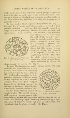
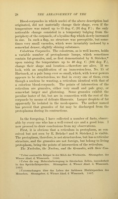
![;i liiitip. Sdiiic of the lumps rciiiiiiii in tlie sliai)e described; most of tlu'in, however, iifter Ji few iiiiiiutes, j^Tadiially resiinu^ the granular condition with increase of their cii-cninfci-ence; at the same time tliey become gloltnlai- and .luiiiii nucleated. Such an ama'ba remains motionless. In still older infnsions, amoeba> make their appearance which are characterized by large size, slowness of locomotion, and the property of pushing out radiating offshoots. Such amoeba^ are especially sensitive to the action of distilled water. By placing a dro]) of distilled Avater at one edge of the covering- glass, and draining off the infusion water from the opposite edge, the folhnving observation was made: Instead of long, radiating offshoots, broad and short flaps weri^ protruded, the long offshoots already present were gradually retracted, and loco- motion ceased. Slowly the amoeba assumed a blunt, polygonal shape. In its body vacuoles appeared, at first small, gradually largei; the nucleus became indistin(?t, and afterward completely faded away. Some of the granules, with jerking movement, united into lai-ger groups, while others were seen to float in larger meshes. The more the number of such free granules increased, the more the amoeba approached the globular shape. At the same time, many small vacuoles coalesced into a single large one, and a globular protrusion at the periphery of the l)ody resulted. Both the protrusion and the body of the amoel)a were seen to be inclosed by a continuous shining layer. Within the vacuole, small granules moved about. In the meantime the jerking motion of the groiiped granules had ceased, and the number of the floating ones became considerably augmented. This description of the appearances after addition of water is taken from one amoeba; but all amoebge of the radiating variety, under similar circumstances, exhibited the same features. Blood-corpuscles of the ('rfor-fish (Astacns). In a drop of blood, transferred from a broken limb of a living fresh craw-fish upon a slide, and covered with a covering-glass oiled on its edges, we recognize the l)lood-corpuseles with moderate powers of the microscope. E. Haeckel * found these corpuscles to be amo-boid. Two kinds of such corpuscles are noticeable, \\z.: pale and finely granular ones with nuclei, which are either large and pale or small and coarsely granular; and, second, others having only coarse, yellowish, very shining granules. The granules of the latter kind, as a rule, encircle light spaces containing scanty granules. * Ueber die Gewebe des Flusskrebses. Miiller's Archiv. 1857.](https://iiif.wellcomecollection.org/image/b21219163_0049.jp2/full/800%2C/0/default.jpg)
 No text description is available for this image
 No text description is available for this image
 No text description is available for this image