Microscopical morphology of the animal body in health and disease / by C. Heitzmann. With 380 original engravings.
- Carl Heitzmann
- Date:
- 1883
Licence: Public Domain Mark
Credit: Microscopical morphology of the animal body in health and disease / by C. Heitzmann. With 380 original engravings. Source: Wellcome Collection.
Provider: This material has been provided by the Augustus C. Long Health Sciences Library at Columbia University and Columbia University Libraries/Information Services, through the Medical Heritage Library. The original may be consulted at the the Augustus C. Long Health Sciences Library at Columbia University and Columbia University.
51/884 page 25
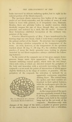
![exposed to (lnylijj:ht, tlu'yellow, sliiniiij^-suhstaiiee wliieli pi-odiiees the reticulum in the i)r(»t()i)l;isiu and its nucleus assumed a vaolct coloi', while the su])stance within the meshes remained nncolored. * li/ood of fill' Xiirt (TritonJ. In a dro}) of l)lood of the newt, transferred to the glass slide and covered with a <'overing-glass oiled on its edges, we can observe tlu' motion of the coloi-less })h)od-corpuscles for hours at the temperature of the room. The changes of shape result from a })rotrusion of light flaps and granular offslioots of varying l)i-cadth, and sometimes of con- siderable length, from the periphery of the protoi)lasmic l)ody. Many finely granular l>odies exhi})it in their interior a con- tinuous change of the grouping of the grannies. Especially after addition of a one-half ])er cent, solution of chloride of sodium, during the locomotions of the body, vacuoles arise, and the inner surface of the wall of many vacuoles looks jagged, as if beset with torn points. For moments the whole body is vacuoled, the smallest, just percei)tible, vacuoles l)eing transitions to still smaller meshes, the filaments of w'liich show pale gray granules as points of intersection ; this appearance is only tem- porary, as the next moment most, or even all, of the vacuoles may have disappeared. In fresh blood, some coarsely granular colorless blood-cor- puscles distinctly show filaments emanating from the gi*anules. With the assistance of an immersion-lens, Xo. 13 of Hartuack, I became con\dnced that, during the locomotion of the corpuscle, the single granules continually keep changing their size and shape, as well as their location. In the blood of newts which had been kept aU winter, the nuclei of many colored blood-corpuscles were ^isil)le, ])oth imme- diately after the mounting of the specimen and after it had remained a time on the glass slide. Each nucleus exhibits a number of coarse, very shining granules, some of which show filaments that are united with the neigh])oring granules. The nucleus is bordered by a continuous layer of a sul>stance of the .same refraction and color as the granules. There is often seen around the nucleus a very narrow light rim, which at times * According to N. Lieberkiibn (Uebor Bewegungserseheiiiimgen der Zelleu, 1870), in blood-corpuscles of many caterpillars thei'e exists a space between the contractile layer and the nucleus which is traversed by fila- ments extending from the inner surface of the contractile layer to the outer surface of the nucleus.](https://iiif.wellcomecollection.org/image/b21219163_0051.jp2/full/800%2C/0/default.jpg)