Microscopical morphology of the animal body in health and disease / by C. Heitzmann. With 380 original engravings.
- Carl Heitzmann
- Date:
- 1883
Licence: Public Domain Mark
Credit: Microscopical morphology of the animal body in health and disease / by C. Heitzmann. With 380 original engravings. Source: Wellcome Collection.
Provider: This material has been provided by the Augustus C. Long Health Sciences Library at Columbia University and Columbia University Libraries/Information Services, through the Medical Heritage Library. The original may be consulted at the the Augustus C. Long Health Sciences Library at Columbia University and Columbia University.
67/884 page 41
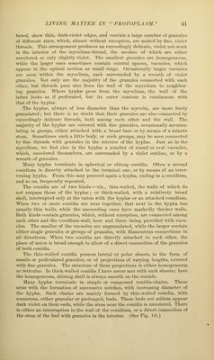
![berod, show thin, (hirk-viok't cdf^os, and coiitaiii a hirj^o Tiurnbcr of {^•aiiuh's of difftMHMit sizes, which, aiiiiost without oxc'<<])ti()ii, am united by fine, viohft tlireads. This urnin^i'intMit produces an exccediiif,dy delicate, violet net-work in the interior of the niyceliuin-tliread, the meshes of which are either uncolored or only slij^htly violet. The smallest {j^anules are homogeneous, while the larger ones sometimes contain central spaces, vacuoles, which appear in the optical section as small rings. Occasioiuilly larger vacuoles are seen within the mycelium, each surrounded by a wreath of violet granules. Not only are the majority of the granules connected with each other, but threads pass also from the wall of the mycelium to neighbor- ing granules. Wliere hyphro grow from the mycelium, the wall of the latter looks as if perforated, but its outer contour is continuous with that of the hypha^ The hyplue, always of less diameter than the mycelia, are more finely gi-anulated; but there is no doubt that their gi-anules are also connected by exceedingly delicate threads, both among each other and the wall. The majority of the hypha^ are covered with fine graimles, occasionally accumu- lating in gioups, either attaclied with a broad base or by means of a minute stem. Sometimes such a little body, or such gi'oups, may be seen coimected by fine threads with gi'anules in the interior of the hyphaj. Just as in the mycelium, we find also in the hyphaj a number of round or oval vacuoles, which, uncolored themselves, are surrounded by a violet outline, or by a wreath of gi'anules. Many hyphie terminate in spherical or oblong conidia. Often a second conidium is directly attached to the terminal one, or by means of an inter- vening hypha. From this may proceed again a hypha, ending in a conidium, and so on, frequently repeated. The conidia are of two kinds — viz., thin-walled, the walls of which do not sm-pass those of the hypha?; or thick-walled, with a relatively broad shell, interrupted only at the union with the hyphae or an attached conidium. When two or more conidia are near together, that next to the hypha has usually thin walls, while the succeeding ones have markedly thicker walls. Both kinds contain granules, which, without exception, are connected among each other and the conidium-wall, here and there being provided with vacu- oles. The smaller of the vacuoles are ungranulated, while the lai'ger contain either single granules or groups of granules, with filamentous connections in all directions. ^V^leIl two conidia are directly attached to each other, the place of union is broad enough to allow of a dii-ect connection of the granules of both conidia. The thin-walled conidia possess lateral or polar shoots, in the form of sessile or pediculated gi'ainiles, or of projections of varying lengths, covered with fine gi'anules. The structure of these projections is either homogeneous or reticular. In thick-walled conidia I have never met with such shoots; here the homogeneous, shining shell is always smooth on the outside. Many hypha; terminate in simple or compound conidia-chains. These arise with the formation of successive notches, with increasing diameter of the hypha. Such chains are mainly formed by thin-walled conidia, with numerous, either granular or prolonged, buds. These buds not seldom appear dark violet on their ends, while the stem near the conidia is uncolored. There is either an iuteiTuption in the wall of the conidium, or a direct connection of the stem of the bud with granules in the interior. (See Fig. 10.)](https://iiif.wellcomecollection.org/image/b21219163_0067.jp2/full/800%2C/0/default.jpg)