Microscopical morphology of the animal body in health and disease / by C. Heitzmann. With 380 original engravings.
- Carl Heitzmann
- Date:
- 1883
Licence: Public Domain Mark
Credit: Microscopical morphology of the animal body in health and disease / by C. Heitzmann. With 380 original engravings. Source: Wellcome Collection.
Provider: This material has been provided by the Augustus C. Long Health Sciences Library at Columbia University and Columbia University Libraries/Information Services, through the Medical Heritage Library. The original may be consulted at the the Augustus C. Long Health Sciences Library at Columbia University and Columbia University.
69/884 page 43
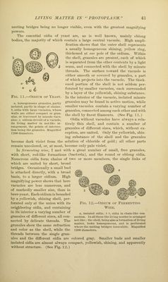
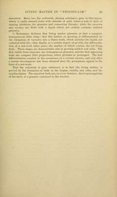
![nj €/ Fig. 1 1.—OiDiUM of Yeast. a, hoiUKgoiieous fp-anulcs, partly isolated, partlj- in shape of cliaius ; 6, oitlia with larfre vacuoles, whose walls arc either coiiipa<-t or gran- uhir, or traversed by minute vacu- oles ; c, oidium devoid of a vacuole, Its living matter in net-like ar- rangement, the points of intersec- tion being the granules. Magnified 1200 diameters. nectinj; bridges bi'iiif^ no Uirif^cr visililc, even with tlie j^reatest iriaf^iifyiiig powers. Tile essential oidiii of yeust are, as is well known, mainly oblong bodies, the majority of which contain a large central vacuole. High ampli- lication shows that the outer shell represents a usually homogeneous shining, yellow ring, thickened at one pole of the oidium. Within the shell, granules are ])resent, each of which is separated from the other contents by a light seam, and connected with the shell by minute threads. The siu'faee toward the vacuole is either smooth or covered Vjy granules, a part of which projects into the vacuole. The thick- ened portion of the shell is not seldom per- forated by smaller vacuoles, each surrounded by a layer of the yellowish, shining substance. In the interior of the vacuole, isolated minute granules may be found in active motion, while smaller vacuoles contain a varying number of gi'anules, connected among each other and with the shell by finest filaments. (See Fig. 11.) Oidia vrithout vacuoles have always a rela- tively thin shell, and contain a number of granules of different sizes, which, without ex- ception, ai*e united. Only the yellowish, shin- ing substance of the shell and the granules become dark violet from the solution of chloride of gold; all other parts remain uncolored, or, at most, become only pale violet. In fcrmentitKi wine, I met with a great number of small, free granules, with short, rod-like formations (bacteria), and the round or oblong oidia. Numerous oidia form chains of two or more members, the single links of which are united by short, broad bridges. Occasionally a small bud is attached tlirectly, with a broad basis, to a larger oidium. High magnifying power shows that here vacuoles are less numerous, and of markecUy smaller size, than in beer-yeast. Each oidium is bounded by a yelloAvish, shining shell, per- forated only at the union with its neighboring oidia, and containing in its interior a varjing number of granules of different sizes, all con- nected by delicate threads. The granules show the same refraction and color as the shell, while the threads between the single gi'an- ules and the different oidia are colored gray. Smaller buds and smaller isolated oidia are almost always compact, yellowish, shining, and apparently without structirre. (See Fig. 12.) Fig. 12.—OiDirM of Wine. Fermenting a, isolated oidia; b h, oidia in chain-like con- nection. In all these the living matter is arranged net-like; the shell, being also a formation of living matter, looks homogeneous, and is perforated where the uniting bridges inosculate. Magnified 1200 diameters.](https://iiif.wellcomecollection.org/image/b21219163_0069.jp2/full/800%2C/0/default.jpg)
 No text description is available for this image
 No text description is available for this image
 No text description is available for this image