Microscopical morphology of the animal body in health and disease / by C. Heitzmann. With 380 original engravings.
- Carl Heitzmann
- Date:
- 1883
Licence: Public Domain Mark
Credit: Microscopical morphology of the animal body in health and disease / by C. Heitzmann. With 380 original engravings. Source: Wellcome Collection.
Provider: This material has been provided by the Augustus C. Long Health Sciences Library at Columbia University and Columbia University Libraries/Information Services, through the Medical Heritage Library. The original may be consulted at the the Augustus C. Long Health Sciences Library at Columbia University and Columbia University.
77/884 page 51
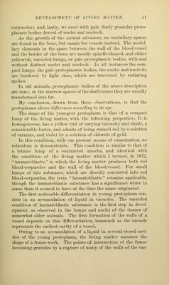
![corpuscU's ; and. lastly, we iiu'ct with pale, finely jifranular proto- plasmic bodies devoid of nuelei and nucleoli. As the growth of the animal advances, no medullary spaces are found in the ])one, but canals for vessels instead. The medul- lary elements in the space Itetween the wall of the ])lood-vessel and the border of the l)one are mostly spindle-slia])ed, and either yello\\'ish, vacuoled lumps, or pale protoplasmic bodies, with and without distinct nuclei and nucleoli. In all instances the com- pact lum})s, the ])ale ])rotoplasmic bodies, the nuclei and nucleoli, are bordered by light rims, which are traversed by radiating spokes. In old aninuils. pi-otoplasmic bodies of the above description ai-e rare; in the nuirrow spaces of the shaft-bones they are usually transformed into fat. My conclusion, drawn from these observations, is that the proiophismn shows difftreiires (icronliny to its (ujc The shape of the youngest protoplasm is that of a compact lump of the li\'ing matter, \A'ith the following properties: It is homogeneous, has a yellow tint of varpng intensity and shade, a considerable luster, and admits of being stained red by a solution of carmine, and violet by a solution of chloride of gold. In this condition, with our present means of examination, no reticulum is demonstrable. This condition is similar to that of a tetanic lump of a contracted amceba, and identical w4th the condition of the living matter which I termed, in 1872, *' ha?matol)lastie, in which the li^'ing matter produces both red blood-corpuscles and the wall of the blood-vessel. For small lumps of this substance, which are directly converted into red blood-corpuscles, the term hfematoblastic'' remains appUcable, though the hfematoblastic substance has a significance wider in sense than it seemed to have at the time the name originated. The first noticeable differentiation in young protoplasm con- sists in an accumulation of liquid in vacuoles. The vacuoled condition of haematoblastic sul)stance is the first step in devel- opment, as observed in the lumps and nuclei of the tissues of somewhat older animals. The first formation of the walls of a vessel depends on this differentiation, inasmuch as the vacuole represents the earliest ca\dty of a vessel. Owing to an aceiunulation of a liquid in several closed cavi- ties of the young protoplasm, the Li\dng matter assumes the shape of a frame-work. The points of intersection of the frame becoming granules by a rupture of many of the walls of the vac-](https://iiif.wellcomecollection.org/image/b21219163_0077.jp2/full/800%2C/0/default.jpg)