Microscopical morphology of the animal body in health and disease / by C. Heitzmann. With 380 original engravings.
- Carl Heitzmann
- Date:
- 1883
Licence: Public Domain Mark
Credit: Microscopical morphology of the animal body in health and disease / by C. Heitzmann. With 380 original engravings. Source: Wellcome Collection.
Provider: This material has been provided by the Augustus C. Long Health Sciences Library at Columbia University and Columbia University Libraries/Information Services, through the Medical Heritage Library. The original may be consulted at the the Augustus C. Long Health Sciences Library at Columbia University and Columbia University.
81/884 page 55
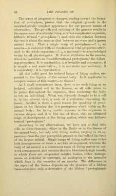
![Till' size of a living- Ixxly is not included in the definition of an or<>^anized individual. In the infusion, f. i., we see g^rowingf g7'aniiles, just percepti])le to the highest inao;nifying powers of the mieroseope, in a fluid in which none were seen a short time before.- The smallest individuals which we are ca])able of seeing with the best mici-oscopes of to-day, are granules; but we must admit that germs or particles of living matter may be present in the air or in fluids in infinite numbers, which cannot be seen at all, and l)econu' visil)le only after having attained a certain size. How comj)licated the structure of a minute particle of living matter may be, we can hardly imagine ; what we do know is, that the so-called '' cell is composed of innumerable })artieles of living matter, every one of which is endowed with properties formerly attributed to the cell-organism. The observati(m of the phases of development of the living matter demonstrates that the term ''cell was attached to only a limited number of forms, diu'ing the changes that take jilace in a growing granule of a substance known to be the seat of life. As the term protoplasm was adapted to the original idea of the cell, it also meant only one or a few phases in the development of a lump of living matter. (See Fig. 19.) Fig. 19.—Diagram of the Phases of Development of the Living Matter. L, series of developiiient of a small granule, a, into a vacuoled lum]), h and c, and into a frame-work, d. P, series of development into jjrotoplasm of a reticular structure; tlie so-called •' cell, e, -n-ith a solid, /, </, li, with vacuoled nuclei. B, series of development tending toward the formation of basis-substance; in i, the nucleus reticular, the nucleolus solid; In k and I, the nucleolus splitting; and in m, the original granule a transformed into a fiuelj-reticular mass, destitute of nucleus and nucleolus.](https://iiif.wellcomecollection.org/image/b21219163_0081.jp2/full/800%2C/0/default.jpg)
 No text description is available for this image
 No text description is available for this image
 No text description is available for this image