Microscopical morphology of the animal body in health and disease / by C. Heitzmann. With 380 original engravings.
- Carl Heitzmann
- Date:
- 1883
Licence: Public Domain Mark
Credit: Microscopical morphology of the animal body in health and disease / by C. Heitzmann. With 380 original engravings. Source: Wellcome Collection.
Provider: This material has been provided by the Augustus C. Long Health Sciences Library at Columbia University and Columbia University Libraries/Information Services, through the Medical Heritage Library. The original may be consulted at the the Augustus C. Long Health Sciences Library at Columbia University and Columbia University.
95/884 page 69
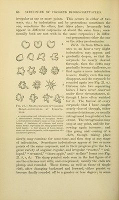
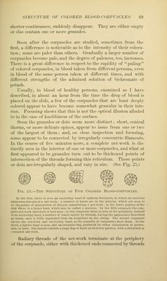
![sThTcrriiH or coi.oiiFjt lu.ooD-coin'rscLKs. m shorter coiitinuaiKH', suddenly dis;i])i»('ar. Tlit^y are either empty or else eoutaiii one oi- iiioi*e ^rautdes. Soon after the eoi'pnscles are studied, sometimes from the first, a difference is noticeable as to the intensity of their colora- tion ; some are paler tlian others. Gradually a larger number of corpuscles become pale, and the degree of paleness, too, increases. There is a great difference in respect to the rapidity of paling of colored corpuscles, in blood taken from different persons, even in blood of the same person taken at different times, and with different strengths of the admixed solution of bichromate of potash. Usually, in l)lo()d of healthy persons, examined as I have described, in about an hour from the time the drop of blood is placed on the slide, a few of the corpuscles that are least deeply colored appear to have become somewhat granular in their inte- rior. Focusing shows that this is notthe optical illusion alluded to in the case of knobbiness of the siu'face. Soon the granules or dots seem more distinct; short, conical thorns, or more delicate spines, appear to issue from one or two of the largest of them; and, on close inspection and focusing, some appear to be connected by ii-regularly concentric filaments. In the course of five minutes more, a complete net-work is dis- tinctly seen in the interior of one or more corpuscles, and what at first appeared to he granules turn out to be thickened points of intersection of the threads forming this reticulum. These points or dots are irregularly shaped, and vary in size. (See Fig. 2).) Fig. 2.).—The Structure of Five Colored Blood-corpuscles. In the liist, tliere is seen an ciiciiclinir hand of unifovm tliickncss, in wliicli are inserted nnnierons tliroails of a network; a nninlxT of knots are in tlie interior, wliicli are seen to 1)0 the points of intersection of tlireails eonstitntinfr a net-work: in tlie lower portion of the ilisk there is a larjrer knot, which may be called a unelens. In the fifth coriiiiscle the com- plete net-work strneture is best seen ; in this cori'nscle there is seen at the periphery, instead of an encircling hand, a number of knots united by threads, having the appearance described a.s beads, each a little separated from its neighlxn-s on the string. Tlie second corjiuscle shows the net-work and encircling band, as the nia.lorit.v of corpuscles show them. In the third, a lighter baud is seen, and an irregular tlaj), produced by either indentation or protru- sion, or both. The fourth exhibits a large flap or knob at its lower portion, with a stretched or extended net-work. Radiary threads of the net-work terminate at the periphery of the corpuscle, either with thickened ends connected by threads](https://iiif.wellcomecollection.org/image/b21219163_0095.jp2/full/800%2C/0/default.jpg)