The surgical treatment of the diseases of infancy and childhood / by T. Holmes.
- Timothy Holmes
- Date:
- 1868
Licence: Public Domain Mark
Credit: The surgical treatment of the diseases of infancy and childhood / by T. Holmes. Source: Wellcome Collection.
435/700
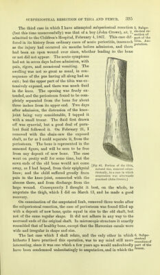
![The third case in which I have attempted subperiosteal resection (but this time unsuccessfully) was that of a boy (John Crowe), set. 7, admitted to the Children's Hospital, February 4,1867. This case dif- fered in its history from ordinary cases of acute periostitis, inasmuch as the injury had occurred six months before admission, and there had been an open wound ever since, whether leading to the bone or not did not appear. The acute symptoms had set in seven days before admission, with pain, rigors, and occasional vomiting. The swelling was not so great as usual, in con- sequence of the pus having all along had an exit; but the upper part of the tibia was ex- tensively exposed, and there was much fluid in the knee. The opening was freely ex- tended, and the periosteum found to be com- pletely separated from the bone for about three inches from its upper end. Two days after admission, the distension of the knee- joint being very considerable, I tapped it with a small trocar. The fluid first drawn off was synovial, but a good deal of puru- lent fluid followed it. On February 21, I removed with the chain-saw the exposed shaft, as far as I could separate it, from the periosteum. The bone is represented in the annexed figure, and will be seen to be free from any deposit of new bone. The case went on pretty well for some time, but the sawn ends of the old bone would not come away, as I had hoped, from their epiphysial lines; and the child suffered greatly from pain in the knee-joint, connected with the abscess there, and from discharge from the large wound. Consequently I thought it best, on the whole, to amputate the thigh, which I did on March 13, and he made a good recovery. On examination of the amputated limb, removed three weeks after the subperiosteal resection, the case of periosteum was found filled up with a deposit of new bone, quite equal in size to the old shaft, but not of the same regular shape. It did not adhere in any way to the necrosed ends of the original shaft. In microscopic structure it exactly resembled that of healthy bone, except that the Haversian canals were wide and irregular in shape and size. The last case which I shall relate, and the only other in which hitherto I have practised this operation, was to my mind still more interesting, since it was one which a few years ago would undoubtedly have been condemned unhesitatingly to amputation, and in which the 5. Subpe- riosteal re- section of part of the tibia. Am- putation. [Fig. 63. Portion of the tibia, natural size, removed subpe- riosteally, in a case in which amputation was afterwards practised (John Crowe).] 6. Subpe- riosteal resection of part of the femur.](https://iiif.wellcomecollection.org/image/b20416325_0435.jp2/full/800%2C/0/default.jpg)