The surgical treatment of the diseases of infancy and childhood / by T. Holmes.
- Timothy Holmes
- Date:
- 1868
Licence: Public Domain Mark
Credit: The surgical treatment of the diseases of infancy and childhood / by T. Holmes. Source: Wellcome Collection.
449/700 page 407
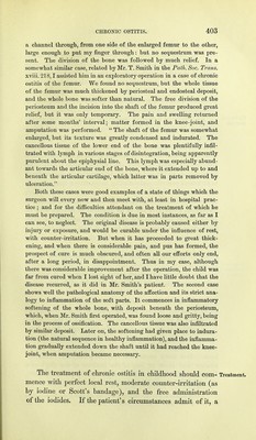
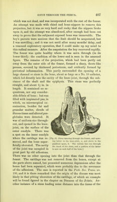
![which was not dead, and was incorporated with the rest of the femur. An attempt was made with chisel and bone-nippers to remove this projection, but it was so very hard and rocky that the nippers broke upon it, and the attempt was abandoned, after enough had been cut away to prove that the subjacent exposed bone was immovable. The boy's parents were anxious that the limb should be amputated, but I was unwilling; and it was not until after many months' delay, and a renewed exploratory operation, that I could make up my mind to this radical measure. After the amputation the boy recovered rapidly. The femur was quite healthy where it was sawn through (in its lower third) : the condition of the lower end is seen in the annexed figure. The remains of the projection, which had been partly cut away from the outer side of the femur, formed a sharp, thorn-like process, covered by thickened periosteum, and evidently formed by periosteal inflammation. This partly surrounded the opening of a large channel or sinus in the bone, about as large as a No. 10 catheter, which led directly into the cavity of the knee-joint, through the sub- stance of the shaft and the epiphysis. This sinus was perfectly straight, and about 1£ in. in length. It contained no se- questrum, nor any consider- able debris of bone; but was filled with inspissated pus, in which, on microscopical ex- amination, besides fat and granular matter, shreds of fibrous tissue and altered pus- globules were detected. It was of uniform size through- out, and opened in the knee- joint, on the surface of the outer condyle. There was a spot on the inner condyle where the cartilage was re- [Mg. 65. Sinus running through the femur, and open- moved and the bone super- ne'ovv ™ *'le knee-joint, a, and above in the - . . x i mi popliteal space, b. The section has run through ficially ulcerated. The cavity the track of the sinuSj and a porti(m of the latter of the joint was occupied in is seen on each half of the bone.] great part by old adhesions. There was no other opening into the joint than that through the femur. The cartilage was not removed from the bones, except at the parts above named, but presented numerous depressions after the bones had been separated, which were probably due to the severance of the adhesions. The case is reported in the Path. Soc. Trans, xv. 190, and it is there remarked that the origin of the disease was most likely in that pitting ulceration of the cartilage, of which an example will be found figured in the chapter on Diseases of the Joints. An- other instance of a sinus leading some distance into the tissue of the](https://iiif.wellcomecollection.org/image/b20416325_0449.jp2/full/800%2C/0/default.jpg)