Licence: Public Domain Mark
Credit: Studies of the internal anatomy of the face / by M.H. Cryer. Source: Wellcome Collection.
Provider: This material has been provided by the Augustus C. Long Health Sciences Library at Columbia University and Columbia University Libraries/Information Services, through the Medical Heritage Library. The original may be consulted at the the Augustus C. Long Health Sciences Library at Columbia University and Columbia University.
18/200
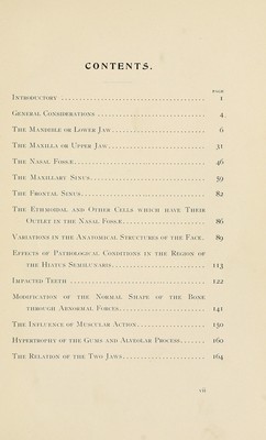
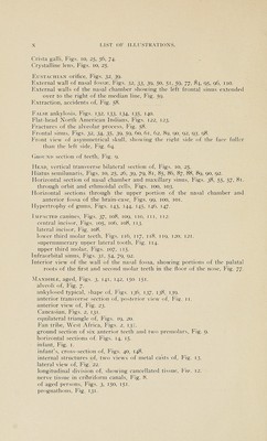
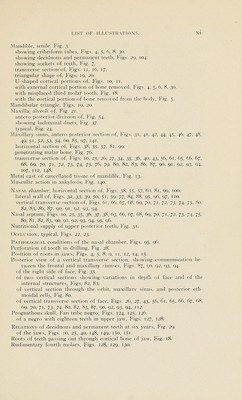
![Section showing diseased condition of inner wall of orbit, Fig. 102. showing greater portion of the upper jaw. Fig. y], through the maxillo-malar articulation. Fig. 76. Separating infraorbital sinus. Fig. 31. Skull, adult. Figs. 22, 23, 24, 150, 151. asymmetrical. Figs. 64, 65. of a fully developed embryo cut vertically through the first deciduous molars, Figs. 40, 148. of aged person. Figs. 150, 151. of Caucasian, Figs. 124, 125, 126. of Fan tribe negro. West Africa. Figs. 124, 125, 126. of Flat-head Indian, Figs. 122, 123. of six-year-old child. Figs. 29, 104. prognathous, Figs. 124, 126, 127. typical, Figs. 22, 23, 24. Sockets of the teeth. Figs. 7, 21. Sphenoidal sinus. Figs. 39, 51, 63, 84. Supernumerary teeth. Figs. 105, 114. Teeth, impacted. Figs, zi, 105, 106, 107, 108. 109, no, in, 112, 113, 114, 115, 116, 117, 118, 119, 120, 121. . rudimentary, Figs. 127, 128, 129, 130. supernumerary. Figs. 105, 114. Temporo-mandibular articulation, modification of. Figs. 141, 142. Transverse section of face. Figs. 10, 25, 26, 27, 34, 35, 36. 40, 43, 56, 61, 65, 66, 67, 68, 69, 70, 71. 72, 73, 74, 75, 79, 80. 82, 83, 86, 87, 90, 91, 92, 93, 94, 107, 112, 148, 149. True ankylosis, mandibles of. Figs. 136, 137. skulls of. Figs. 138, 139. Two antero-posterior sections made by dividing the orbit and maxillary sinus vertically, showing conical elevations over the roots of the various teeth. Fig. 41. Two illustrations showing variations in the depth and size of the external walls of the nasal chambers. Fig. 84. Two sections showing diseased condition of ethmoidal cells, Fig. 103. Under surface of two upper jaws, showing comparison in size, Fig. 129. Upper and lower jaws, anterior view of, Fig. 149. ' Upper jaw, anterior lateral view of. Figs. 4, 30. anterior view of, Fig. yi- antero-posterior section of. Figs. 31, 41, 42, 45, 46, 47, 48, 49, 52, 53, 54, 97. showing deciduous and permanent teeth. Fig. 29. transverse section of. Figs. 10, 25; 26, 27, 40, 43, 56. under surface and alveoli of the various teeth. Fig. 21. tmder surface of, showing occluding surfaces of teeth. Figs. 24, 126, 128, 129, 130. vertical transverse section of, showing ostium maxillare and infraorbital sinus, Fig. 79. Vertical antero-posterior division through the frontal sinus, orbit, and max- illary sinvis. Fig. 44. Vertical transverse section of asymmetrical skull. Figs. 65, 66, 67, 68, 69, 70, 71.](https://iiif.wellcomecollection.org/image/b21225096_0018.jp2/full/800%2C/0/default.jpg)


 No text description is available for this image
 No text description is available for this image
 No text description is available for this image