Licence: Public Domain Mark
Credit: A textbook of physiology / By M. Foster. Source: Wellcome Collection.
Provider: This material has been provided by the Augustus C. Long Health Sciences Library at Columbia University and Columbia University Libraries/Information Services, through the Medical Heritage Library. The original may be consulted at the the Augustus C. Long Health Sciences Library at Columbia University and Columbia University.
29/1072
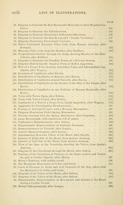
![PAGE [152 Cerebro-spinal Axis, showing Origin of Cranial and Spinal Nerres, after Marshall, 626] [153. Various Forms of Ganglionic Vesicles, after Kirke, 628] [154. Structure of Pyrifonu Nerve-cells, after Beale and Arnold, . , . 629] [155. Stellate Nerve-cell, after Beale 629] [156. Funiculus of Nerve, after Ranvier, 630] [157. Nerve-fibre from Sciatic Nerve, after Ranvier, 630] [158. Nerve-tubules showing Effects of Post-mortem Changes, after Wagner, 631] [159. Diagram of Structure of Nerve-fibre, after Carpenter, .... 631] [160. Piimitive Nerve-fibrils, after Schultze, 632] [161. Diagram of a Horizontal Section of the Eyeball, after Ellis, . . . 645] [162. Vertical Section of Cornea, alter Ellis, 646] [163. Inner View of the Front of the Choroid Coat, the Ciliary Processes, and the Back of the Iris, after Ellis, 647] [164. Section of the Ciliary Region of the Eye of Man, after Iwauoff, . . 648] [165. Muscular Structure of the Iris, after KoUiker, 647] [166. Pigment-cells of the Eyeball, after Kolliker, 650] [167. Diagrammatic Representation of the Connection of the Nerve-fibres of the Retina, after Schultze, 651] [168. Rod and Cone from the Retina of Man, after schultze 651] [169. Diagrammatic Representation of the Connective Tissue of the Retina, after Schultze, ......... ^ •. ■ 651] [170. Inner Surface of the Retina, showing Macula Lutea, Entrance of Optic Nerve, and Arteria Centralis Retina, after Soemmering, . . . . 653] [171. Magnified Vertical Seciion of Retina, after Ellis, 653] [172. Representation of the Laminge of a Hardened Lens, after Ellis, . . 654] 173. Diagram of Scheiner's Experiment, 660 [174. Diagram showing the Course of Parallel Rays in the Myopic Eye, after Carpenter, 661] [175. Diagram showing the Course of Parallel Rays in the Hypermetropic Eye, after Carpenter, 662] [176. Diagram showing the Course of Parallel Rays in the Emmetropic Eye, after Carpenter, 662] [177. Diagram showing how the Accommodation of the Lens is affected for Di- verging Rays of Near Objects, after Carpenter, ...... 664] 178. Diagram illustrating Chromatic Aberration, 675 [179. Diagram of Method for Finding the Situation of the Blind Spot, . . 678] 180. Diagram illustrating the Formation of Purkinje's Figures when the Illu- mination is directed through the Sclerotic, 680 181. Diagram illustrating the Formation of Purkiuje's Figures when the Illu- mination is directed through the Cornea, 680 182. Diagram of the Three Primary Color Sensations 698 183. Diagram illustrating the Appreciation of Apparent Size, .... 708 184. Diagram illustrating Corresponding Points in the Retina, . . . .710 185. Diagram of the Attachment of the Muscles of the Eye, after Frick, . 714 186. Diagram illustrating a Simple Horopter, 718 187. Diagram illustrating Monocular and Binocular Perception of an Object, . 721 [188. Vertical Section of the Meatus Auditorius Externus and Tympanum, after Scarpa, 726] [189. Inner View of Membrana Tympani and Tympanic Bone, after Ellis, . 728] [190. Plan of Ossicles in Position in the Tympanum, with their Muscles, from Ellis, 728]](https://iiif.wellcomecollection.org/image/b21222149_0029.jp2/full/800%2C/0/default.jpg)



 No text description is available for this image
 No text description is available for this image
 No text description is available for this image