Licence: Public Domain Mark
Credit: A textbook of physiology / By M. Foster. Source: Wellcome Collection.
Provider: This material has been provided by the Augustus C. Long Health Sciences Library at Columbia University and Columbia University Libraries/Information Services, through the Medical Heritage Library. The original may be consulted at the the Augustus C. Long Health Sciences Library at Columbia University and Columbia University.
31/1072
![PAGE 223. Side and Upper Views of the Brain of Man, with the Areas of the Cere- bral Convolutions, according to Ferrier, 831 224. Side and Upper Views of the Brain of ^[an, with the Areas of the Cere- bral Convolutions, according to Ferrier, 832 [225. Course of Fibres in the Optic Commissure, after Bowman, . . . . 861] [226. The Nerves of the Orbit seen from the Outer Side, after Arnold, . . 8G2J [227. General Plan of the Branches of the Fifth Pair, after Charles B.^11,. . 86o] [228. The Distribution of the Facial Nerve, after Erasmus Wilson, . . . 867] [229. The Distribution of the Pneumogastric Nerve, after Hirschftld and Le- veille 870] [230. Median Section of the Mouth, Nose, Pharynx, and Larynx, after Quain, 874] [231. View of the Larynx and part of the Trachea behind, after Quain, . . 87G] [232. View of the Larynx from above, after Quain, 876] [233. View of Upper Part of the Larynx, as seen by means of tiie Laryngo- scope, during the Utterance of a Grave Note, after Quain, . . . 876] 234. The Larynx as seen by means of the Laryngoscope in Different Condi- tions of the Glottis, from Quain, after Czermak, 878 [235. Illustration of Third Kind of Lever, after Kirke, 889] [236. Diagram illustrating Movements of Legs and Feet in Walking, after Kirke, .... - 890] [237. Diagrammatic View of the Uterus and its Appendages, after Quain, . 895] [238. Section of the Liuins: Membrane of Uterus, after Weber, .... 896] [239. View of a Section of a Prepared Ovary of a Cat, after Sehron, . . . 897] [240. The Base of the Male Bladder, with VesiculieSeminales and Prostate Gland, after Ilaller, 898] [241. View of a Section of the Testicle and Epididymis, after Kirke, . . 898] [242. Spermatozoa and Spermatic Ceils, after Kolliker, 900] [243. Section of a Graafian Follicle, after Vcn Baer 901] [244. Ovum of a Sow, after Barry, 901] [245. Successive Stages of the Formation of the Corpus Luteum, after Pouchet, 903] [246. Diagram of the Various Stages of the Cleavage of the Yolk, after Dalton, 907] [247. Impregnated Egg, with the Commencement of the Formation of the Em- bryo, after Dalton 908] [248. Diagrammatic Section of Ovum, at Fifteenth or Seventeenth Day, . . 909] [249. Diagrammatic Section of Ovum, with Chorion and Villi, after Todd and Bowman, 910] [250. Diagrammatic Section of Ovum, with Chorion and Villi, after Todd and Bowman, 910] [251. Diagram of Fecundated Egg, after Dalton, 911] [252. Diagramof Fecundated Egg, with Aliantois nearly complete, after Dalton, 911] [253. Human Ovum, at eighth week,showing Tufts of the Chorion, after Ecker, 912] [254. Portion of one of the Foetal Villi, after Ecker, 912] [255. First Stage of the Formation of the Decidua Refiexa, after Coste, . . 913] [256. More Advanced Stagp of the Decidua Reflexa, after Coste, .... 913] [257. Section of a Fully Formed Placenta, after Ecker, 914] [258. Diagram of the Foetal Circulation, after Erasmus Wilson, .... 921] [259. Cbolesterii., after Dalton 988]](https://iiif.wellcomecollection.org/image/b21222149_0031.jp2/full/800%2C/0/default.jpg)

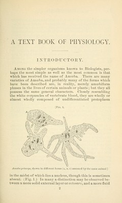


 No text description is available for this image
 No text description is available for this image
 No text description is available for this image