Elements of comparative anatomy / by Carl Gegenbaur ; translated by F. Jeffrey Bell ; the translation revised and a preface written by E. Ray Lankester.
- Gegenbaur, C. (Carl), 1826-1903.
- Date:
- 1878
Licence: Public Domain Mark
Credit: Elements of comparative anatomy / by Carl Gegenbaur ; translated by F. Jeffrey Bell ; the translation revised and a preface written by E. Ray Lankester. Source: Wellcome Collection.
640/686 (page 606)
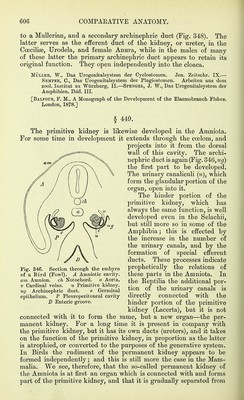
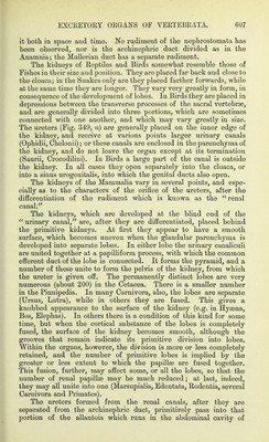
![to a MuUeriaiij and a secondary archinepliric duct (Fig. 348). The latter serves as the efferent duct o£ the kidney, or ureter, in the Coecilias, Urodela, and female Anura, while in the males of many of these latter the primary archinephric duct appears to retain its original function. They open independently into the cloaca. MÜLLER, W., Das Urogenitalsystem der Cyolostomen. Jen. Zeitsclix'. IX.— Semper, C, Das Urogenitalsystem der Plagiostomen. Arbeiten aus dem zool, Institut zu Würzburg, II,—Spengel, J, W,, Das Urogenitalsystem der Amphibien. Ibid. III. [Balfour, F. M,, A Monograph of the Development of the Elasmobranch Fishes. London, 1878.] § 449. The primitive kidney is likewise developed in the Amniota. For some time in development it extends through the coelom, and projects into it from the dorsal wall of this cavity. The archi- nephric duct is again (Fig. 346,iig) the first part to be developed. The urinary canaliculi (u), which form the glandular portion of the organ, open into it. The hinder portion of the primitive kidney, which has always the same function, is well developed even in the Selachii, but still more so in some of the Amphibia; this is effected by the increase in the number of the urinary canals, and by the formation of special efferent ducts. These processes indicate prophetically the relations of these parts in the Amniota. In the Reptilia the additional por- tion of the urinary canals is directly connected with the hinder portion of the primitive kidney (Lacerta), but it is not connected with it to form the same, but a new organ—the per- manent kidney. For a long time it is present in company with the primitive kidney, but it has its own ducts (ureters), and it takes on the function of the primitive kidney, in proportion as the latter is atrophied, or converted to the purposes of the generative system. In Birds the rudiment of the permanent kidney appears to be formed independently; and this is still more the case in the Mam- malia. We see, therefore, that the so-called permanent kidney of the Amniota is at first an organ which is connected with and forms part of the primitive kidney, and that it is gradually separated from Fig. 346. Section through the embyro of a Bird (Fowl). A Amniotic cavit5^ am Amnion. ch Notochord. a Aorla. V Cardinal veins. u Primitive kidney. ug Archinephric duct. e Germinal epithelium. P Pleuroperitoneal cavity D Enteric groove.](https://iiif.wellcomecollection.org/image/b20417202_0640.jp2/full/800%2C/0/default.jpg)