Licence: Public Domain Mark
Credit: Selected monographs. Source: Wellcome Collection.
Provider: This material has been provided by UCL Library Services. The original may be consulted at UCL (University College London)
361/372 page 57
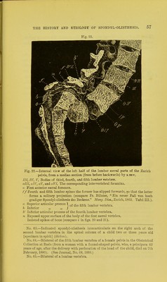
![Fig. 22. Fig. 22.—Internal view of the left half of the lumbar sacral parts of the Zurich pelvis, from a median section (from before backwards) by a saw. Ill, IV, V, Bodies of third, fourth, and fifth lumbar vetebrse. oIII, oIV, oV, and oVI, The corresponding intervertebral foramina. o First anterior sacral foramen. ff Fourth and fifth lumbar spines the former has slipped forward?, so that the latter forms a solitary projection (compare Fr. Billeter, Ein neuer Fall von hoch gradiger Spondylolisthesis des Beckens. Mang. Diss., Zurich, 1862. Tafel III.). a Superior articular process 1 q{ ^ fifth lumbar vertebra. b Inferior „ „ J V Inferior articular process of the fourth lumbar vertebra. n Exposed upper surface of the body of the first sacral vertebra. Isolated spikes of bone (compare i in figs. 20 and 21). No. 63.—Indicated spondylolisthesis interarticularis on the right arch of the second lumbar vertebra in the spinal column of a child two or three years old [specimen in spirit] (ibidem). No. 64.—Bilateral of the fifth lumbar vertebra of a female pelvis in the Obstetrical Collection at Basle (from a woman with a funnel-shaped pelvis, who, a primipara 42 years of age, after the delivery with perforation of the head of the child, died on 7th February, 1880). (See Journal, No. 68, 1880.) No. 65.—Bilateral of a lumbar vertebra.](https://iiif.wellcomecollection.org/image/b21290167_0361.jp2/full/800%2C/0/default.jpg)


 No text description is available for this image
 No text description is available for this image
 No text description is available for this image