On rest and pain : a course of lectures on the influence of mechanical and physiological rest in the treatment of accidents and surgical diseases, and the diagnostic value of pain / delivered at the Royal College of Surgeons of England in the years 1860, 1861, and 1862 by John Hilton.
- John Hilton
- Date:
- 1879
Licence: Public Domain Mark
Credit: On rest and pain : a course of lectures on the influence of mechanical and physiological rest in the treatment of accidents and surgical diseases, and the diagnostic value of pain / delivered at the Royal College of Surgeons of England in the years 1860, 1861, and 1862 by John Hilton. Source: Wellcome Collection.
Provider: This material has been provided by the Harvey Cushing/John Hay Whitney Medical Library at Yale University, through the Medical Heritage Library. The original may be consulted at the Harvey Cushing/John Hay Whitney Medical Library at Yale University.
29/320
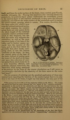
![itself, and lines the under surface of the brain, crura cerebri, pons Varolii, medulla oblongata, &c. Tracing this membrane (the internal arachnoid) posteriorly, its'posterior layer passes underneath the cerebellum, joined to the internal laver of theexternal arachnoid; it then quits the external arachnoid, and follows the under surface of the cerebellum until it reaches the cerebro-spinal opening, which it enters. You notice, therefore, that this large space (d), which I now term the internal arachnoid cavity, and which contains the cerebro- spinal fluid, not only passes down the whole length of the vertebral canal (the relative position is indi- cated in this diagram), but the fluid lies under and supports the cerebellum, as well as the most important parts of the base of the brain; and it further passes up- wards into the interior of the brain, and is the fluid which we generally find, after death, occu- pying more or less the various ventricles of the brain. In fact, the central parts of the base of the brain, instead of resting upon the bones of the skull at its base, rest upon this collection of cere- bro-spinal fluid, which forms for it a most beautiful, efficient, and perfectly adapted water-bed; the water-bed itself being sustained in its position by the force of the venous circulation—as I will prove to you presently—and also by the elasticity of the dura mater in the verte- bral canal.* For the purpose of pointing out the practical application of these ana- tomical facts regarding the function of the cerebro-spinal fluid in its rela- tion to the base of the brain, I might say that almost the only part of the Fig. 5.—n, Cnt extremity of medulla. Immediate- ly in front of this is the large space occupied by cerebro- spinal fluid, b b. Under surface of cerebellum, c c, Middle lobes, d d. Anterior lobes. * This passage has been left as it was originally written by Mr. Hilton. The reader will see at once that the space between the two layers of the external arachnoid M corresponds to the cavity of the arachnoid, and that the interval between the two layers of the internal arachnoid v is the sub-arachnoid space between the arachnoid and the pia matsr. Mr. Hilton believes that the cerebro-spinal fluid is contained between two epithelial surfaces, i e., that one such surface not only exists, as usually described, upon the arachnoid, but also upon the pia mater as well, and that thus the otherwise inevitable infiltration of the pia mater is prevented. With all due deference to Mr. Hilton. I cannot quite accept this view. In the first place, as far as I am able to make out microscopic n 11 y and with the use of such reagents as silver nitrate, no layer of epithelial cells exists upon the pia mater. I am further inclined to believe that a slight infiltration of the superficial part of the pia mater does take place, as the cerebro-spinal fluid passes backwards and forwards over its surface. More than very slight infiltration is prevented, partly by the fact that the cerebro-spinal fluid is broken up into a number of little lakes and pools by the very delicate bands which are found in the sub-arachnoid space (the tissue forming these bands being also infiltrated by the fluid), and partly by the deeper layers being most closely united to the brain substance by the innumerable vessels entering it. I am indebted to Mr Davies-Colley for the knowledge of the fact that Henle applies to the sub-arachnoid tissue the epithet wassersiichtig, cedematous. — [Ed.]](https://iiif.wellcomecollection.org/image/b2102005x_0029.jp2/full/800%2C/0/default.jpg)