The oriental sore, as observed in India : a report / by T.R. Lewis and D.D. Cunningham.
- Timothy Richards Lewis
- Date:
- 1877
Licence: Public Domain Mark
Credit: The oriental sore, as observed in India : a report / by T.R. Lewis and D.D. Cunningham. Source: Wellcome Collection.
58/74 page 48
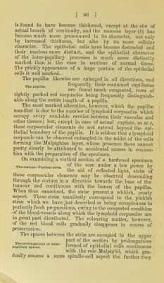
![[ ] The vascular structures. of both classes of follicles were frequently infiltrated with accumulations of lymphoid cells. The sweat glands, situated in the lower part of the Th, .udorip.ro„,gl„d.. P; /’ appreciably aftected unless the sore is in a very advanced state. Frequently the glandular con- volutions may be seen to present an almost perfectly normal appearance (Fig. 7, r), even when surrounded by the lym- phoid corpuscles. The ducts, however, are generally larger than usual, and sometimes they are seen to have become obliterated, apparently as the result of the pressure exercised by the newly-formed cells of the sore. Occasionally, how- ever, the appearance presented by the cellular lining of the ducts and its convolutions seemed to indicate that, as in the case of the sebaceous glands, some change had occurred, but whether neoplastic or retrograde we were unable to decide. It is in connection with the vascular structures of the corium that the essential features of the disease are most prominently observed. This, however, does not imply that the primary deposit takes place in the interior of the vessels, or even in the interior of the capillaries ; for very frequently sections of the blood-vessels parallel to their course may be encountered, which prove that they contain no corpuscular elements other than those normal to them, although it is frequently to be noted that they are very full. The same is observable in transverse sections. The coats of the vessels are, however, generally thickened and somewhat more readily resolved into their cellular elements than is the case in normal tissues. In endeavouring to trace a connection between the lymphoid cells and the various tissues of the corium, all preparations de- monstrate more or less clearly that the distribution of these elements is more intimately connected with the delicate fibrous tissue- investment of the glandular and vascular structures than with any other tissue. In following the course of a blood- vessel, for example, it will be observed that aggregations— colonies of lymphoid cells—have formed in numerous places, and that from these “ colonies ” ragged processes of delicate fibrous tissue may be recognised which become joined to simi- lar processes in other parts of the preparation (Fig. 7, h,h,h). In other instances these delicate fibrous shreds may be seen to be directly continuous with the adventilia of a vessel, and Relation of the lymphoid corpuscles to the fibrous tissue-investments of vessels and glands.](https://iiif.wellcomecollection.org/image/b28709615_0058.jp2/full/800%2C/0/default.jpg)