Histology; normal and morbid.
- Dunham, Edward K. (Edward Kellogg), 1860-1922
- Date:
- 1898
Licence: Public Domain Mark
Credit: Histology; normal and morbid. Source: Wellcome Collection.
Provider: This material has been provided by the Augustus C. Long Health Sciences Library at Columbia University and Columbia University Libraries/Information Services, through the Medical Heritage Library. The original may be consulted at the the Augustus C. Long Health Sciences Library at Columbia University and Columbia University.
107/466 page 111
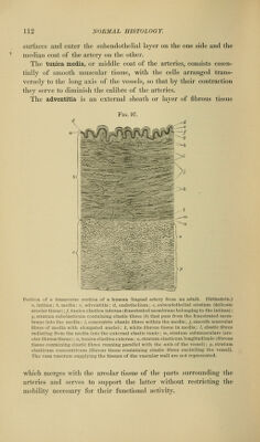
![the vessel is contracted they are thicker and the })ortion containing the nucleus projects slightly into the lumen of the vessel. The subendothelial fil)rous tissue forming the second layer of the intima is composed of very delicate fibrils, closely packed together, with a little cement l)etween them, and enclosing irregular spaces in which the iji-anching cells of the tissue lie. Elastic fibres, spring- FiG. 96. Branch of splenic artery of a rabbit: o, internal endothelial surface of the intima; 6, elastic lamina of the intima (fenestrated membrane, see Fig. 59); c, media composed of smooth muscular tissue encircling the vessel and therefore appearing in longitudinal section with elongated nuclei: d, adventitia of fibrous tissue blending above and to the left with the surrounding areolar tissue; e. adipose tissue, between the cells of which a few lines of red corpuscles reveal the presence of capillary bloodvessels; /, small nerve, containing both medullated and pale or non-medullated nerve-fibres. There are other similar sections of nerves in the figure. To the left of the artery the section is slightly torn, the adipose tissue being separated from the adventitia of the artery. A few red blood-corpuscles have been extravasated near the nerve at the upper left corner of the figure. There are also a few corpuscles within the lumen of the arterj-. ing from the external layer of the intima, may here and there, especially in the larger arteries, make their way into the subendo- thelial layer. The clastic lamina of the intima is formed by a network of anas- tomosing elastic fibres, having a general longitudinal disposition with respect to the axis of the vessel. The spaces left between the fibres of this network vary considerably in size. Where they are small and the fibres between them are correspondingly broad this layer has the a])pearance of a perforated membrane (the fenestrated mem- brane of Henle). Even where this membranous character of the elastic layer is well developed, elastic fibres are given off from its](https://iiif.wellcomecollection.org/image/b21223841_0107.jp2/full/800%2C/0/default.jpg)