Histology; normal and morbid.
- Dunham, Edward K. (Edward Kellogg), 1860-1922
- Date:
- 1898
Licence: Public Domain Mark
Credit: Histology; normal and morbid. Source: Wellcome Collection.
Provider: This material has been provided by the Augustus C. Long Health Sciences Library at Columbia University and Columbia University Libraries/Information Services, through the Medical Heritage Library. The original may be consulted at the the Augustus C. Long Health Sciences Library at Columbia University and Columbia University.
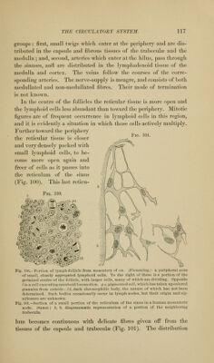
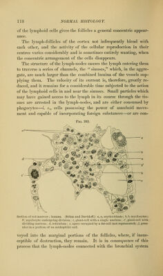
113/466 page 117
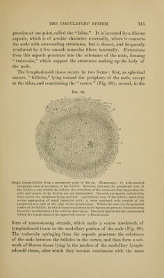
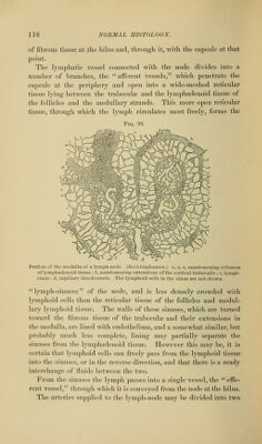
![p;roup«;: first, small t\vi<;s wliicli enter at the ])eripherv and are dis- tril)nte(l in the eapsnle and til)rons tissncs of the trabecular and the medulla ; and, second, arteries which enter at the hilus, pass through the sinuses, an7l are distributed in the lyniphadenoid tissue of the medulla and cortex. The veins follow the courses of the corre- sponding arteries. The nerve-supply is meagre, and consists of both medullated and non-meduUated fibres. Their mode of termination is not known. In the centre of the follicles the reticular tissue is more open and the lymphoid cells less abundant than toward the periphery. Mitotic figures are of fre(juent occurrence in lymphoid cells in this region, and it is evidently a situation in which those cells actively multiply. Further toward the periphery the reticular tissue is closer and very densely packed with small lymphoid cells, to be- come more open again and freer of cells as it passes into the reticulum of the sinus (Fig. 100). This last reticu- FiG. 100. Fio. 101. @/?i) ^^'y^ mmMm Fig. 100.-Portion of lympli-foUiclc from mesentery of ox. (Flemming.) z. peripheral zone of small, closely apffreRated lymphoid cells. To the right of these is a portion of the germinal centre of the follicle, with larger cells, many of which are dividing. Opposite / is a cell executing amceboid locomotion, p z, pigmented cell, which has taken up colored granules from oiitside: tk, dark chromophilic body, the nature of which has not been determined. Such bodies occasionally occur in lymph-nodes, but their origin and sig- nificance are unknown. Fig. 101.—Section of a small portion of the reticulum of the sinus in a human mesenteric node. (Saxer.) b, b, diagrammatic representation of a portion of the neighboring trabecula. lum becomes continuous Avith delicate fibres given off from the tissues of the capsule and trabeculae (Fig. 101). The distribution](https://iiif.wellcomecollection.org/image/b21223841_0113.jp2/full/800%2C/0/default.jpg)