Histology; normal and morbid.
- Dunham, Edward K. (Edward Kellogg), 1860-1922
- Date:
- 1898
Licence: Public Domain Mark
Credit: Histology; normal and morbid. Source: Wellcome Collection.
Provider: This material has been provided by the Augustus C. Long Health Sciences Library at Columbia University and Columbia University Libraries/Information Services, through the Medical Heritage Library. The original may be consulted at the the Augustus C. Long Health Sciences Library at Columbia University and Columbia University.
127/466 page 131
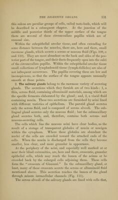
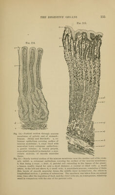
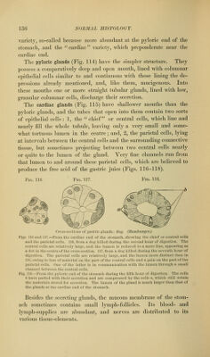
![this sulcus are peculiar groups of colls, called taste-buds, which will he described in a subsequent chapter. At the junction of the middle and posterior thirds of the upper surface of the tongue there are several of tiiese circumvallate papillae which are of muisual size. Within the subepithelial areolar tissue, and often extending for some distance between the muscles, there are, here and there, small racemose glands, which secrete a serous or mucous fluid (Figs. 109, a and 110). They are most abundant on the back and sides of the pos- terior part of the tongue, and their ducts frequently open into the sulci of the circumvallate papilla\ Within the subepithelial areolar tissue small collections of lymphadenoid tissue (lymph-follicles) are also of not infrequent occurrence. The papillae covering these are low and inconspicuous, so that the surface of the tongue appears unusually smooth at those points. 2. The salivary glands belong to the racemose variety of secreting glands. The secretions which they furnish are of two kinds : 1, a thin, serous fluid, containing albuminoid materials, among which are the specific ferments elaborated by the gland; and, 2, a viscid fluid containing mucin. These two secretions are furnished by acini lined with different varieties of epithelium. The parotid gland secretes onlv the serous fluid, and is composed of serous alveoli. The sub- liuffual g-land secretes onlv the mucous fluid ; but the submaxillar^' gland secretes both, and, therefore, contains both serous- and mucous-secreting cells. The cells which line the mucous acini have clear bodies, as the result of a storage of trans])arcnt globules of mucin or mucigen within the cytoplasm. Where these globules are abundant the nuclei of the cells are crowded toward the attached ends of the cells. When the mucin is discharged from the cells they become smaller, less clear, and more granular in appearance. At the periphery of the acini, and especially well marked at or near their blind extremities, are, here and there, crescentic, granular epithelial cells, which may reach the lumen of the acinus or be crowded back by the enlarged cells adjoining them. These cells form the crescents of Gianuzzi. In the submaxillary gland, at least, many of these crescents secrete the serous or albuminoid fluid mentioned above. This secretion reaches the lumen of the gland through minute intracellular channels (Fig. 111). The serous alveoli of the salivary glands are lined M-ith cells that,](https://iiif.wellcomecollection.org/image/b21223841_0127.jp2/full/800%2C/0/default.jpg)