Histology; normal and morbid.
- Dunham, Edward K. (Edward Kellogg), 1860-1922
- Date:
- 1898
Licence: Public Domain Mark
Credit: Histology; normal and morbid. Source: Wellcome Collection.
Provider: This material has been provided by the Augustus C. Long Health Sciences Library at Columbia University and Columbia University Libraries/Information Services, through the Medical Heritage Library. The original may be consulted at the the Augustus C. Long Health Sciences Library at Columbia University and Columbia University.
172/466 page 176
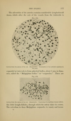
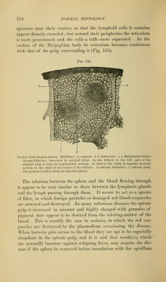
![CHAPTER XIV. THE SPLEEN. Nearly the whole surface of the spleen is invested with a cov- ering of peritoneum similar to that which partially covers the liver. Beneath this is the true capsule of the spleen, which com- pletely surrounds it. This capsule is composed of dense fibrous tissue, containing a large number of elastic fibres and a few of smooth muscular tissue. From its inner surface bands of the same tissue, called the trabeculse, penetrate into the substance of the organ, M^iere they branch, and the branches join each other to form a coarse meshwork occupied by the parenchyma of the organ, the '' pulp. The bloodvessels of the spleen enter at the hilum and pass into the large trabeculse, which start from the capsule at that point and enclose the vessels until they divide into small branches. The vessels then leave the trabeculse and penetrate the pulp, where they break up into capillaries, which do not anastomose with each other. There is some doubt as to the way in which these capillaries end. According to one view, they unite to form the venous radicles, so that the blood is confined within vessels throughout its course in the spleen. Another view, which is more probably correct, is that the walls of the capillaries become incomplete, clefts appearing between tlieir endothelial cells, which finally change their form and become similar to those of the reticulum of the pulp. The veins, accord- ing to this view, arise in a manner similar to the endings of the arteries. The result of this structure would be that the blood is discharged, from the capillary terminations of the arteries, directly into tlie meshes of the pulp, after which it is taken up by the capillary origins of the veins (Figs. 151 and 152). The pulp consists of a fine reticulum of delicate fibres and cells, with branching and communicating ])rocesses, in the meshes of wliich there are red blood-corpuscles, leucocytes in greater number than nonually present in the blood, and free amoeboid cells consid- eral)ly larger than leucocytes, called the splenic cells.](https://iiif.wellcomecollection.org/image/b21223841_0172.jp2/full/800%2C/0/default.jpg)