Histology; normal and morbid.
- Dunham, Edward K. (Edward Kellogg), 1860-1922
- Date:
- 1898
Licence: Public Domain Mark
Credit: Histology; normal and morbid. Source: Wellcome Collection.
Provider: This material has been provided by the Augustus C. Long Health Sciences Library at Columbia University and Columbia University Libraries/Information Services, through the Medical Heritage Library. The original may be consulted at the the Augustus C. Long Health Sciences Library at Columbia University and Columbia University.
185/466 page 189
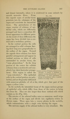
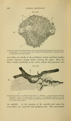
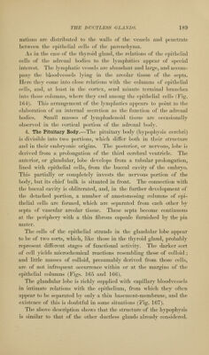
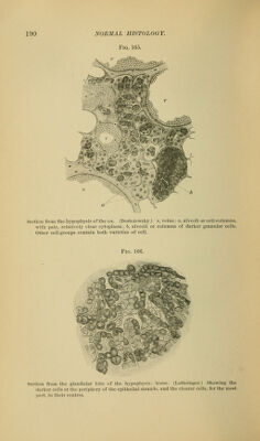
![nations arc distributed to the wails of tiie vessels and penetrate between the epithelial cells of the parenchyma. As in the ease of the thyroi<l gland, the relations of the epithelial cells of the adrenal bodies to the lynij)hatics apj)ear of special interest. The lymphatic vessels are abundant and large, and accom- pany the bl(Jodvessels lying in the areolar tissue of the septa. Here they come into close relations with the columns of epithelial cells, and, at least in the cortex, send minute terminal branches into those columns, where they end among the epithelial cells (Fig. 104). This arrangement of the lym})hatics appears to point to tlx; elaboration of an internal secretion as the function of the adrenal bodies. Small masses of lymphadenoid tissue are occasionally observed in the cortical [)ortion of the adrenal bodv. 4. The Pituitary Body.—The pituitary body (hypophysis cerebri) is divisible into two portions, which differ both in their structure and in their eml)ryonic origins. The posterior, or nervous, lobe is derived from a prolongation of the third cerebral ventricle. The anterior, or glandular, lobe develops from a tubular prolongation, lined with epithelial cells, from the buccal cavity of the embryo. This partially or completely invests the nervous portion of the body, but its chief bulk is situated in front. The connection with the buccal cavity is obliterated, and, in the further development of the detached ])ortion, a number of anastomosing columns of epi- thelial cells are formed, which are separated from each other by septa of vascular areolar tissue. These septa become continuous at the periphery with a thin fibrous capsule furnished by the pia mater. The cells of the epithelial strands in the glandular lobe appear to be of two sorts, which, like those in the thyroid gland, probably represent different stages of functional activity. The darker sort of cell yields microchemical reactions resembling those of colloid ; and little masses of colloid, presumably derived from those cells, are of not infre([ucnt occurrence within or at the margins of the epithelial columns (Figs. 165 and 166). The glandular lobe is richly supplied with capillary bloodvessels in intimate relations with the epithelium, from which they often appear to be separated by only a thin basement-membrane, and the existence of this is doubtful in some situations (Fig. 167). The above description shows that the structure of the hypophysis is similar to that of the other ductless glands alreadv considered.](https://iiif.wellcomecollection.org/image/b21223841_0185.jp2/full/800%2C/0/default.jpg)