Histology; normal and morbid.
- Dunham, Edward K. (Edward Kellogg), 1860-1922
- Date:
- 1898
Licence: Public Domain Mark
Credit: Histology; normal and morbid. Source: Wellcome Collection.
Provider: This material has been provided by the Augustus C. Long Health Sciences Library at Columbia University and Columbia University Libraries/Information Services, through the Medical Heritage Library. The original may be consulted at the the Augustus C. Long Health Sciences Library at Columbia University and Columbia University.
334/466 page 338
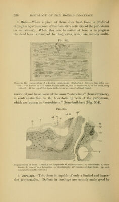
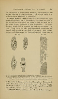
![4. Bone.—Wlien a piece of boue dies fresh bone is produced through a rejuvenescence of the formative activities of the periosteum (or endosteura). While this new formation of bone is in progress tlie dead bone is removed by phagocj'tes, which are usually multi- FiG. 303. Phase in the regeneration of a tendon; guinea-pig. (Enderlen.) Seventy days after sec- tion. The tendon is still rather highly cellular, but its structure is, in the main, fully restored. At the top of the figure is the cross-section of a blood-vessel. nucleated, and have received the name '' osteoclasts (bone-breakers), in contradistinction to the bone-forming cells of the periosteum, which are known as osteoblasts (bone-builders) (Fig. 304). Fig. 304. t\k 'f? nh t ff Ih •fc H III , Ik sp Regeneration of bone. (Barth.) nk, fragments of necrotic bone; rz, osteoclasts; o, osteo- blasts ; Ik, bone of new formation : y, bloodvessels ; nk', lamina of dead bone, (sp, acci- dental crack in the section.) 5. Cartilage.—This tissue is ca])ablc of only a limited and imper- fect regeneration. Defects in cartilage are usually made good by](https://iiif.wellcomecollection.org/image/b21223841_0334.jp2/full/800%2C/0/default.jpg)