Histology; normal and morbid.
- Dunham, Edward K. (Edward Kellogg), 1860-1922
- Date:
- 1898
Licence: Public Domain Mark
Credit: Histology; normal and morbid. Source: Wellcome Collection.
Provider: This material has been provided by the Augustus C. Long Health Sciences Library at Columbia University and Columbia University Libraries/Information Services, through the Medical Heritage Library. The original may be consulted at the the Augustus C. Long Health Sciences Library at Columbia University and Columbia University.
390/466 page 394
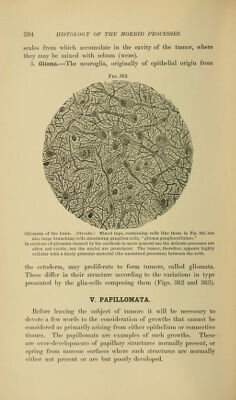
![scales from which accumulate in the cavity of the tumor, where they may be mixed with sebum (wens). 5. Grlioma.—The neuroglia, originally of epithelial origin from Fig. 363. Gliomata of the brain. (Stroebe.) Mixed type, containing cells like those in Fig. 362, but also large branching cells simulating ganglion-cells, glioma gangliocellulare. In sections of gliomata stained by the methods in more general use the delicate processes are often not visible, but the nuclei are prominent. The tumor, therefore, appears highly cellular with a finely granular material (the unstained processes) between the cells. the ectoderm, may proliferate to form tumors, called gliomata. These diifer in their structure according to the variations in type presented by the glia-cells composing them (Figs. 362 and 363). V. PAPILLOMATA. Before leaving the subject of tumors it will be necessary to devote a few words to the consideration of growths that cannot be considered as primarily arising from either epithelium or connective tissues. The papillomata arc exam])les of such growths. These are over-developments of papillary structures normally present, or spring from mucous surfaces where such structures are normally either not present or are but poorly developed.](https://iiif.wellcomecollection.org/image/b21223841_0390.jp2/full/800%2C/0/default.jpg)