Histology; normal and morbid.
- Dunham, Edward K. (Edward Kellogg), 1860-1922
- Date:
- 1898
Licence: Public Domain Mark
Credit: Histology; normal and morbid. Source: Wellcome Collection.
Provider: This material has been provided by the Augustus C. Long Health Sciences Library at Columbia University and Columbia University Libraries/Information Services, through the Medical Heritage Library. The original may be consulted at the the Augustus C. Long Health Sciences Library at Columbia University and Columbia University.
76/466 page 80
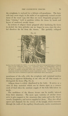
![to the tissue-elements it bathes, and then returns to the blood in the veins through the interstices and lymphatic vessels contained in the areolar tissue. In pursuance of these functions, areolar tissue pervades nearly all parts of the body. Wherever bloodvessels are found, there more or less areolar tissue is present, surrounding them, giving them support, and furnishing channels for the lym- phatic circulation. As has already been stated, this areolar tissue varies in the closeness of its texture in different parts of the body. The fibrous tissues of tendons and ligaments form inextensible Fig. 66. ^m^}^'' Portion of a large tendon in transverse section. (Schafer.) a, sheath of areolar tissue sur- rounding the tendon; 6, longitudinal fasciculus of fibres within that sheath ; I, lymphatic space; c, section of a broad extension of the enshcathing areolar tissue, dividing the tendon into larger bundles; d, e, more delicate layers of areolar tissue subdividing the larger bundles of fibres. Between these areolar septa are the bundles of fibres constitut- ing the tendon. The cells which lie between the smallest fasciculi of fibres appear in stellate form; the cross-sections of the individual fibres, among which these cells lie, are not represented. They would appear as minute dots. Ijands or cords highly resistant to tensile stress, but very ])liable. They consist of bundles of fibres lying parallel to each other and to the direction in which they are to resist pulling forces. Layers of loo.se areolar tissue penetrate the ligaments and tendons, dividing tlif-m into fasciculi, which in turn are united into larger bundles by thicker layers of areolar tissue (Fig. 60). These .sheaths of areolar tissue support the vessels and nerves supplied to the denser forms of the fibrous ti.ssue making up the ligaments or tendons. The thicker aponeuroses of the body may be regarded as broad and flat](https://iiif.wellcomecollection.org/image/b21223841_0076.jp2/full/800%2C/0/default.jpg)