Medical morbid anatomy and pathology / by Hugh Thursfield and William P.S. Branson.
- Thursfield, Hugh.
- Date:
- 1909
Licence: In copyright
Credit: Medical morbid anatomy and pathology / by Hugh Thursfield and William P.S. Branson. Source: Wellcome Collection.
36/302 page 24
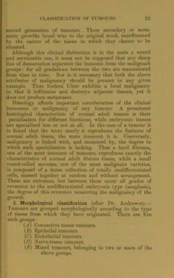
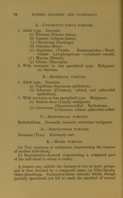
![A.—Connective tissue tumours. 1. Adult type. Innocent. (a) Fibroma (Fibrous tissue). (b) Lipoma (Adipose tissue). (c) Chondroma (Cartilage). (d) Osteoma (Bone). (e) Angeioma (Vessels. Haemangeioma = Blood- vessels. Lymphangeioma = Lymphatic vessels). (/) Myoma (Muscle). (g) Glioma (Neuroglia). 2. With reversion to less specialised type. Malignant. (a) Sarcoma. B.—Epithelial tumours. 1. Adult type. Innocent. (a) Papilloma (Squamous epithelium). (b) Adenoma (Columnar, cubical, and spheroidal epithelium). 2. With reversion to less specialised type. Malignant. (a) Rodent ulcer (Locally malignant). /7, n _ fSquamous-celled. Epithelioma. (<) aicmoma.cu}_>|ca]j spheroidal-celled. C. —Endothelial tumours. Endothelioma. Generally innocent, sometimes malignant. D. —Nerve-tissue tumours. Neuroma (True). Extremely rare. E.—Mined tumours. (a) True teratoma or embryoma (representing the remains of another individual). (b) Sequestration-dermoid (representing a misplaced part of the individual in whom it exists). A tumour may exhibit the features of two or more groups, and is then denoted by a compound name, e.g. Fibro-lipoma, Osteo-chondroma. Connective-tissue tumours which, though partially specialised, yet fail to reach the standard of normal](https://iiif.wellcomecollection.org/image/b2809170x_0036.jp2/full/800%2C/0/default.jpg)
 No text description is available for this image
 No text description is available for this image
 No text description is available for this image