Lectures on ectopic pregnancy and pelvic haematocele / by Lawson Tait, F.R.C.S., Edin., & Eng., LL.D.
- Lawson Tait
- Date:
- 1888
Licence: Public Domain Mark
Credit: Lectures on ectopic pregnancy and pelvic haematocele / by Lawson Tait, F.R.C.S., Edin., & Eng., LL.D. Source: Wellcome Collection.
Provider: This material has been provided by University of Bristol Library. The original may be consulted at University of Bristol Library.
94/122 page 90
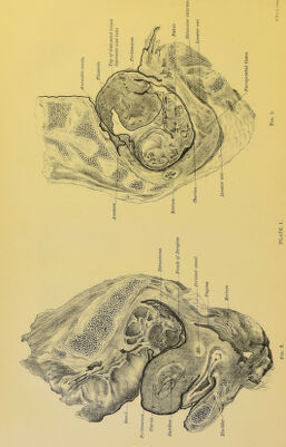
![the anterior abdominal wall and outer surface of peritoneum. Where attached to the anterior abdominal wall, the veins there are enlarged. The diameter of its long axis is 13-5 cm., and its average antero- posterior measurement is 7'5 cm. Around it is a thin investment of connective tissue, and it is firnily attached at points, especially in front and above, to the surrounding capsule by bands of vascu- larized tissue. In the right sections a cavity is seen between the capsule and the placenta, whicli was filled with a mass of grumous blood, and gases of decomposition, the position of whicli corresponds to a well-defined darkening of the skin of the anterior abdominal wall, as if the patient had suffered from a severe blow or fall. The foetus weighed 2 lbs. 4 oz. without the umbilical cord. It was fairly well nourished, hut clccom])osition had commenced. esiKCudly at the lower part of the abdomen. The consideration of these two sections shows, therefore, a special phase in the development of extra-uterine gestation. They demonstrate that a Fallopian tube pregnancy may develop between the layers of the broad ligament, and may continue this extra- peritoneal mode of growth, stripping off the peritoneum from the uterus, bladder, and pelvic floor until it becomes in great part surrounded by a peritoneal capsule derived from these organs. All this is done without any actual intra-peritoneal invasion. The placenta in the advanced gestation case is attached in front to the extra-peritoneal connective tissue, the veins there enlarging and acting like uterine veins. In this special cadaver, therefore, the gestation began probably in the right Fallopian tube, developed into the layers of the broad ligament, and grew extra-peritoneally, lifting up the peritoneum on the right side of the middle line, both anteriorly and posteriorly, and also stripping the posterior uterine wall and upper part of the anterior uterine wall. The extra- peritoneal tissue, with its blood-vessels, is therefore not only capable of forming anastomoses in abdominal aneurism, as Turner and Chiene have shown, but may attempt to carry on the functions of the maternal portion of the placenta. We have liere what may be termed slow displacement of tlie placenta. At first it lay in the Fallopian tube, but the growing ovum has slowly pushed it up (a process attended with blood extravasation) from pelvis to aljdominal cavity, until at last its upper edge is about ten inches from its original site. Part of this is due to growth of course. The uterus also has had its cervical portions elongated in the same way to three inches. These sections have an important bearing on the classification of extra-uterine gestation. AIucli has been written, and little really demonstrated on this point. The Tubal variety is undoubted ; the Tubo-ovarian has also been demonstrated; but the Ovarian is a very doubtful form. The Sub-peritoneo-pelvic or intra-ligamentous variety of](https://iiif.wellcomecollection.org/image/b21448048_0094.jp2/full/800%2C/0/default.jpg)