Mr John Spear's report to the local government board upon the so-called "Woolsorters' Disease" as observed at Bradford and in neighbouring districts in the west Riding of Yorkshire.
- Spear, John.
- Date:
- 1881
Licence: Public Domain Mark
Credit: Mr John Spear's report to the local government board upon the so-called "Woolsorters' Disease" as observed at Bradford and in neighbouring districts in the west Riding of Yorkshire. Source: Wellcome Collection.
Provider: This material has been provided by London School of Hygiene & Tropical Medicine Library & Archives Service. The original may be consulted at London School of Hygiene & Tropical Medicine Library & Archives Service.
36/70 (page 36)
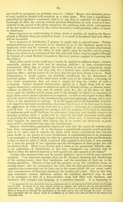
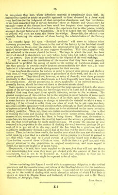
![38 APPENDICES. A. CLINICAL NOTES, NOTES ON AUTOPSIES, OF MICROSCOPICAL EXAMINATIONS AND OF PATHOLOGICAL EXPERIMENTS. [The cases in which Dr. Greenfield carried out pathological experiments are those first recorded here.] (Case 20, for history, page 24) Autopsy.—T. C., aged 71, died 21st May 1880. Autopsy 30 hours after death. Body well developed. Rigor mortis fairly marked, but decomposition considerably advanced. Much dark discolouration of skin (hypostasis and cyanosis), especially of back, arms, neck, and face, which latter was also swollen. A frothy blood-stained fluid issued from mouth and nostrils. Subcutaneous cellular tissue of neck, arms, chest, and, in patches, in various parts of the body and limbs, emphy- sematous ; a crackling sensation being produced on pressure. The epidermis raised in blist rs in some of the more dependent parts, and in moving the body is detached in large strips. Thorax.—Con- siderable jelly-like oedema of the mediastinal connective tissue ; copious straw-coloured serous effusion in pleural cavities ; lungs showing deep hypostatic congestion, and much general oedema. The mucous membrane of the trachea and larger bronchi somewhat hypersemic, and containing some blood- stained frothy mucous ; the bronchial glands swollen and congested. The pericardium contains about 3 ozs. of reddish serum, its surface smooth and lustrous. The heart flaccid, and full of dark cherry- coloured fluid blood, the endocardium being stained w’ith its colouring matter. The abdominal cavity contains a small quantity of blood-stained serous exudation ; the peritoneum injected. The gastric mucous membrane softened and separated in parts from the muscular coat (most marked at fundus and probably p.m.); small intestine in parts much congested, and containing liquid faeces, but no blood ; mesenteric and retro-peritoneal glands enlarged and congested ; the mesentery infiltrated with exudation now gelatinised. The spleen moderately enlarged, dark and pulpy ; kidneys deeply congested, the surrounding connective tissue infiltrated with serum; the liver and other organs apparently normal. Brain not examined. In the blood and other fluids examined immediately after completion of autopsy bacilli were found. Dr. Greenfield’s report is as follows :— “ Case 20. T. C., died May 21, 3 a.m. ; material received May 23rd, 4.30 a.m. Examination made and inoculations performed 5 to 7 a.m., 50 hours after death. Blood from the spleen ; the tissues and T. C.—Blood, showing the bacterial organisms found in it. a. Single and jointed bacilli, several containing spores. b. Long filaments. c. Shorter rods forming groups, of which only a part is represented. d and e. Two of the rods resembling those at a, more highly magnified. This and the other figures, to which a scale is appended, were drawn to scale when fresh, under a magnifying power of 400 diameters, and are represented as enlarged to twice that size. The more highly magnified were drawn under a power of 800 diameters, and are also enlarged to twice natural size. In some of the woodcuts the bacilli are made to appear somewhat too thick. The unit of measurement, /*, a micro-millimetre, equals of-an inch. bloody fluid from the spleen smelt as if decomposed ; they contained a very large number of rods, most of which measured from 5 to 8 yc., but many longer, 10 to 15 //.. The smaller ones resembled both in diameter and shape short rods of bacillus anthracis, but the shorter rods were rounder at the ends than usual. The longer rods were blunt at the ends and contained typical spores. All were motionless. No bacterium termo, but some micrococci. “ The serous fluid from pleura contained many very long rods measuring from 50 to 150 in length, straight or jointed, together with many other shorter rods, in which spores in course of formation or fully formed were visible. “ Sanguineous fluid from the lung also contained abundant long and medium-sized rods of somewhat variable diameter, 1'2 to 1*8 p. “ Inoculations with these fluids were entirely unsuccessful, and need not therefore be detailed.” Fig. 1. (Case 22, page 25.)—Clinical notes by Mr. McKenzie, as follows :— “ Moulson C., mt. 30, a woolsorter, a muscular, athletic, healthy-looking man, who has always previously enjoyed good health. Family history good. States that on the 8th of May 1880 he played at a cricket match and perspired freely, became chilly afterwards, and has not since felt well. He has been sorting Cape hair for the last six months, and feels sure he has got the ‘ sorters disease,’ and it](https://iiif.wellcomecollection.org/image/b24996774_0038.jp2/full/800%2C/0/default.jpg)