Licence: Public Domain Mark
Credit: Human anatomy (Volume 2). Source: Wellcome Collection.
Provider: This material has been provided by the National Library of Medicine (U.S.), through the Medical Heritage Library. The original may be consulted at the National Library of Medicine (U.S.)
13/684
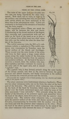
![to commence at the side of the root of the nose by a vein formed by the junction of branches from the forehead, eyebrow, and nose, and to increase by receiving others during its course. The frontal vein, [v. frontalis,] commences on the roof of the skull by branches, which descend obliquely inwards upon the forehead, maintaining communications in their course with the anterior branches of the temporal vein. By gradually converging, these branches form a vein of some size, which descends vertically, parallel with the corresponding vessel of the opposite side, with which it is connected by transverse branches. In some instances the veins of the two sides unite and form a short trunk, which again divides into two branches at the root of the nose. These branches diverge as they run along the sides of the nose at its root, where each becomes continuous with the corresponding angular vein. As it descends from the fore- head, the frontal vein receives a branch from the eyebrow, and some, of smaller size, from the nose and upper eyelid. The supra-orbital vein (v. swpercilii) runs inwards in the direction of the eyebrow, covered by the occipito-frontal muscle. Its branches are connected externally with those of the external palpebral and superficial temporal veins; in its course it receives branches from the contiguous muscles and integument, and at the inner angle of the orbit inclines downwards, to terminate in the frontal vein. The supra-orbital and frontal veins, by their junction, form the angular vein, which is perceptible beneath the skin as it runs obliquely downwards and outwards by the inner margin of the orbit, resting against the side of the nose at its root. This vessel receives by its inner side the nasal veins, which pass upwards obliquely to join it from the side and ridge of the nose; whilst some small palpebral veins open into it from the opposite direction. Opposite the lower margin of the orbit, the angular vein may be said to terminate by becoming continuous with the facial vein. The facial vein, commencing as has been just stated, gradually increases, as it receives branches from the lower eyelid, from the ala of the nose, and from the upper lip. By its outer side it receives two or three veins {inferior palpebral), which are formed by small branches derived from the lower eyelid, from the outer side of the orbit, and from the cheek. The direction of these palpebral branches is obliquely inwards above the zygomatic muscle, beneath which they turn pre- viously to their termination. On a level with the angle of the mouth, the facial vein receives communicating branches (deep facial) from the pterygoid plexus, and also some branches proceeding from the orbit, furnished by the infra-orbital and other branches of the internal maxillary vein. In front, the facial vein is further increased by branches from the lips (labial), and behind by others from the cheek (buccal); still lower down, by branches from the masseter muscle (masseteric) on the one hand, and from the chin on the other. Having reached the base of the lower maxilla, the vein inclines outwards and backwards, covered by the cervical fascia and the platysma muscle; and soon unites with a large branch of communication derived from the temporal vein, to form a vessel of considerable size, which joins obliquely the trunk of the internal jugular vein, k.](https://iiif.wellcomecollection.org/image/b21148879_0013.jp2/full/800%2C/0/default.jpg)