Licence: Public Domain Mark
Credit: The functions of the brain / by David Ferrier. Source: Wellcome Collection.
Provider: This material has been provided by the Royal College of Physicians of Edinburgh. The original may be consulted at the Royal College of Physicians of Edinburgh.
510/528 page 484
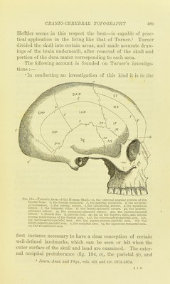
![frontal (f) eminences, and the external angular iDrocess of the frontal bone (a) are easily recognised structures, the position of which can be determined b_y manipulating the scalp, and still more readily on the surface of the skull itself. The coro- nal (c) and lambdoidal (l) sutures can also be felt through the scalp ui most heads, and on the skull itself, the position of the squamous (s), squamoso-sphenoid (ss), and parieto-sphe- noid sutures (j) s), and the curved hne of the temporal ridge (0, can also without difficulty be deternuned ' (Turner, op. eit.) [references inserted]. With these as fixed points the surface of the skull may be divided into ten well-defined areas or regions. The coronal suture (c) forms the posterior boundary of the frontal area. An imaginary hne (fig. 134, (v)) drawn from the squamous suture (s) vertically upwards through the parietal emmence (p) to the sagittal suture or middle line of the skull subdivides the parietal region into an antero-jyarietal (fig. 134, SAP + iAP) and a, post-parietal area (fig. 134, spp-I-ipp). The occipital region, which hes between the lambdoidal suture (I) and the occipital protuberance (o), and the superior curved hne extending on each side from it, forms the occi- pital area (fig. 134, o). These fom- primary divisions are further subdivided. The temporal ridge (fig. 134, t), extending backwards from the external process of the frontal bone (a), across the frontal, antero-parietal, and post-parietal areas to the lateral angle of the occipital bone, di-sddes these areas into an upper and a lower division. We have thus an upper and a lower frontal area (sf and I f) ; an upper antero-parietal (s A p) and a loxcer antero-parietal area (i A p) ; an upper p)ostero-parietal (s p p) and a loicer postero- parictal area (ipp). § 12. The boundaries of these areas are as follows :—The inferior frontal area, or, as it may also be called, the fronto- temporal area, is bounded above by the temporal ridge, below by the fronto-sphenoid suture, and behmd by the coronal suture. The inferior antero-parietal area is bounded above by the temporal ridge, below by the squamous and parieto-sphe- noid sutures, in front by the coronal suture, and behmd by](https://iiif.wellcomecollection.org/image/b21902550_0510.jp2/full/800%2C/0/default.jpg)
 No text description is available for this image
 No text description is available for this image
 No text description is available for this image