A treatise on the diseases of the eye.
- Wells, J. Soelberg (John Soelberg), -1879.
- Date:
- 1873
Licence: Public Domain Mark
Credit: A treatise on the diseases of the eye. Source: Wellcome Collection.
Provider: This material has been provided by the Gerstein Science Information Centre at the University of Toronto, through the Medical Heritage Library. The original may be consulted at the Gerstein Science Information Centre, University of Toronto.
106/868 page 100
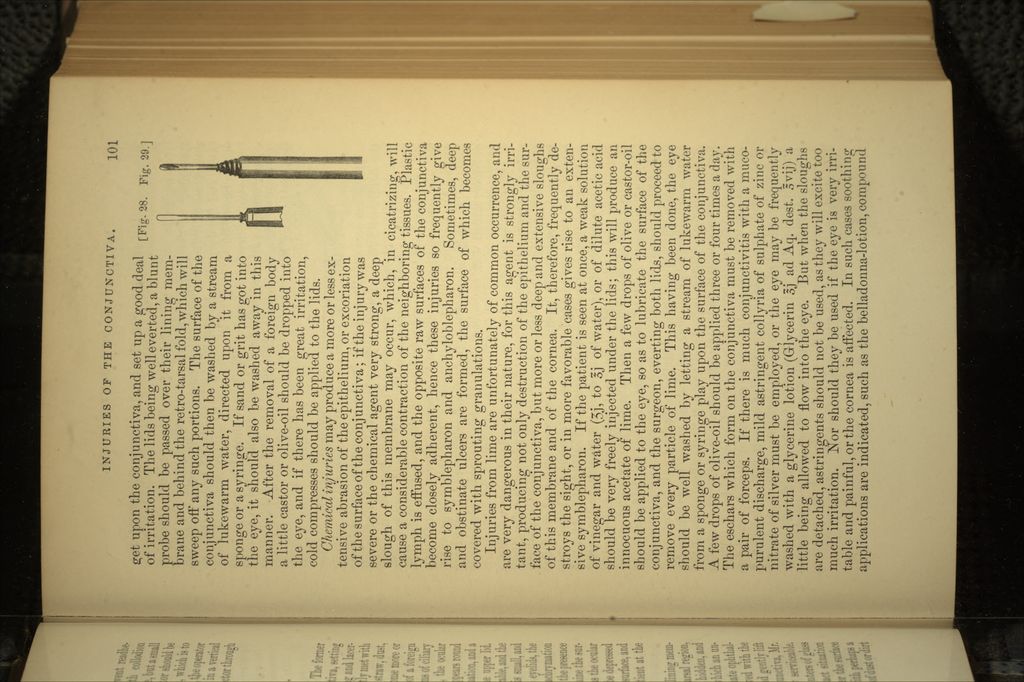
![divided close to the edge of the lid. In order to prevent readhe- sion of the surfaces, these should be touched with collodion (Haynes Walton). If the anchyloblepharon is complete, but a small opening exists near the nasal portion, a grooved director should be passed in through this, and run behind the adhesion, which is to be divided upon it with a scalpel. If no opening exists, the operator should at one point lift up the lids from the eyeball in a vertical fold, and divide the adhesion here, then introduce a director through this incision, and finish the operation with its aid. 14.—INJURIES OF THE CONJUNCTIVA. These may be of a mechanical or chemical nature. The former may prove injurious by their contact with the conjunctiva, setting up irritation and inflammation, or from their wounding and lacer- ating this membrane. The foreign bodies most frequently met with on the conjunctiva are bits of steel, iron, glass, coal, straw, dust, etc., which may remain lodged on its surface, or become more or less deeply imbedded in its structure. The presence of a foreign body in the eye generally sets up at once severe symptoms of ciliary irritation. The eyelids are spasmodically contracted, the ocular conjunctiva becomes injected, and a bright rosy zone appears round the cornea; there is also much photophobia, lachryrnation, and a feeling as of sand and grit in the eye or under the upper lid. Sometimes, the pain and ciliary neuralgia are considerable, and the pupil is markedly contracted. If the foreign body is small, and simply lies on the conjunctiva, the movements of the eyelids, the rubbing of the eye by the patient, and the copious lachrymation will often suffice to extrude it. If the surgeon suspects the presence of a foreign body, he must carefully and closely examine the sur- face of the palpebral conjunctiva of both lids, as well as the ocular conjunctiva and the cornea. The lower eyelid is to be depressed by the fore and middle finger so as to bring its inner surface, and especially the retro-tarsal fold, well into view, the patient at the same time being directed to look upwards. The upper lid is next to be well everted, and its lining mem- brane thoroughly scanned, more particularly the retro-tarsal region, within the folds of which the foreign body often lies hidden, and may easily escape detection. Cases are narrated in which an un- discovered foreign body has set ujp a severe and obstinate ophthal- mia, When found, the foreign body should be removed with the spud [Fig. 28], which should be inserted beneath it, and gently lift it out. If it has got somewhat imbedded in the conjunctiva, Mr. Haynes Walton's gouge [Fig. 29] will be found very serviceable. If the foreign bodies, more especially shot or small splinters of glass or steel, etc., are buried in the conjunctiva, their exact situation should be ascertained by lightly passing the finger over the surface of the conjunctiva, and they should then be excised with perhaps a small portion of the latter. Sometimes, impalpable bits of dust or dirt](https://iiif.wellcomecollection.org/image/b20996408_0106.jp2/full/800%2C/0/default.jpg)
 No text description is available for this image
 No text description is available for this image
 No text description is available for this image