A treatise on the diseases of the eye.
- Wells, J. Soelberg (John Soelberg), -1879.
- Date:
- 1873
Licence: Public Domain Mark
Credit: A treatise on the diseases of the eye. Source: Wellcome Collection.
Provider: This material has been provided by the Gerstein Science Information Centre at the University of Toronto, through the Medical Heritage Library. The original may be consulted at the Gerstein Science Information Centre, University of Toronto.
109/868 page 103
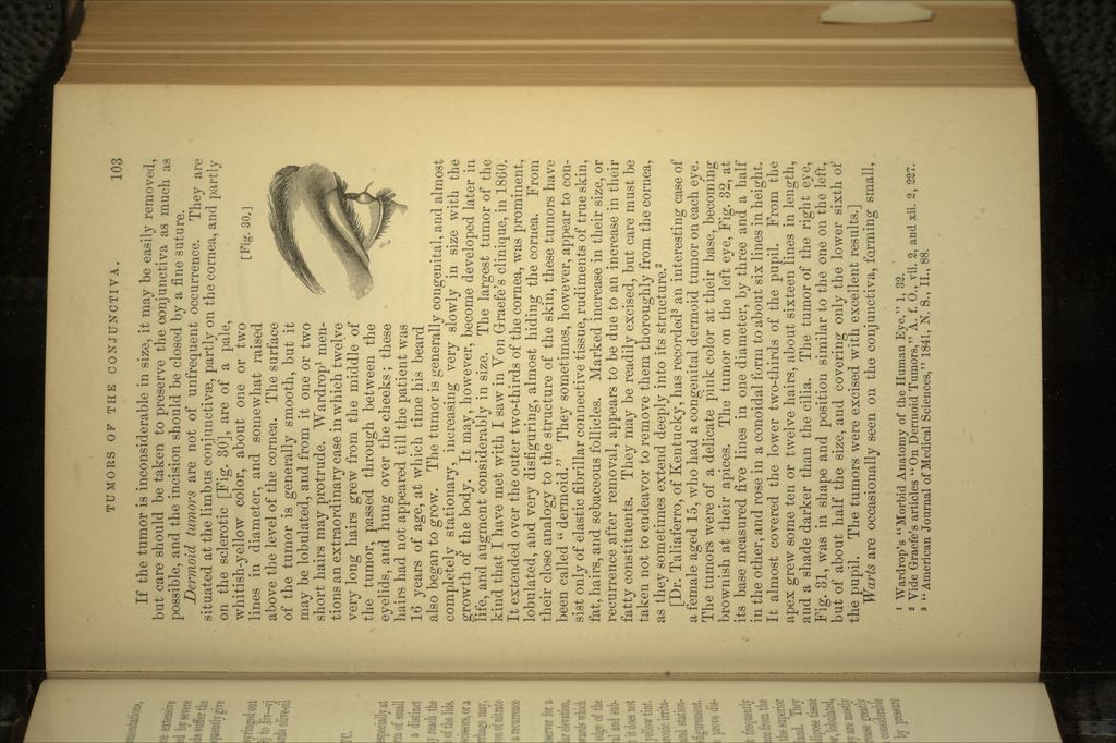
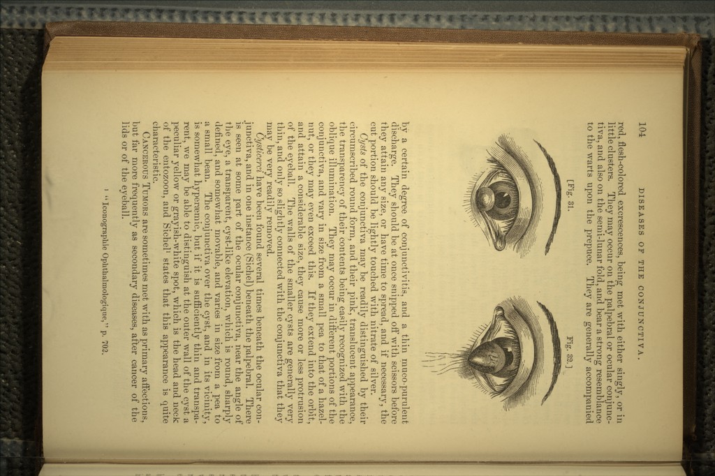
![If the tumor is inconsiderable in size, it may be easily removed but care should be taken to preserve the conjunctiva as much as possible, and the incision should be closed by a fine suture. Dermoid tumors are not of unfrequent occurrence. They are situated at the limbus conjunctiva, partly on the cornea, and partly on the sclerotic [Fig. 30], are of a pale, whitish-yellow color, about one or two [p-lc, 30 -, lines in diameter, and somewhat raised above the level of the cornea. The surface of the tumor is generally smooth, but it may be lobulated, and from it one or two short hairs may protrude. Wardrop1 men- tions an extraordinary case in which twelve very long hairs grew from the middle of the tumor, passed through between the eyelids, and hung over the cheeks; these hairs had not appeared till the patient was 16 years of age, at which time his beard also began to grow. The tumor is generally congenital, and almost completely stationary, increasing very slowly in size with the growth of the body. It may, however, become developed later in life, and augment considerably in size. The largest tumor of the kind that I have met with I saw in Von Graefe's clinique, in 1860. It extended over the outer two-thirds of the cornea, was prominent, lobulated, and very disfiguring, almost hiding the cornea. From their close analogy to the structure of the skin, these tumors have been called dermoid. They sometimes, however, appear to con- sist only of elastic fibrillar connective tissue, rudiments of true skin, fat, hairs, and sebaceous follicles. Marked increase in their size, or recurrence after removal, appears to be due to an increase in their fatty constituents. They may be readily excised, but care must be taken not to endeavor to remove them thoroughly from the cornea, as they sometimes extend deeply into its structure.2 [Dr. Taliaferro, of Kentucky, has recorded3 an interesting case of a female aged 15, who had a congenital dermoid tumor on each eye. The tumors were of a delicate pink color at their base, becoming brownish at their apices. The tumor on the left eye, Fig. 32, at its base measured five lines in one diameter, by three and a half in the other, and rose in a conoidal form to about six lines in height. It almost covered the lower two-thirds of the pupil. From the apex grew some ten or twelve hairs, about sixteen lines in length, and a shade darker than the cilia. The tumor of the right eye, Fig. 31, was in shape and position similar to the one on the left, but of about half the size, and covering only the lower sixth of the pupil. The tumors were excised with excellent results.] Warts are occasionally seen on the conjunctiva, fojming small, 1 Wardrop's Morbid Anatomy of the Human Eye, 1, 32. 2 Vide Graefe's articles On Dermoid Tumors, A. f. 0., vii. 2, and xii. 2, 227. 3 American Journal of Medical Sciences, 1841, N. S., II., 88.](https://iiif.wellcomecollection.org/image/b20996408_0109.jp2/full/800%2C/0/default.jpg)
 No text description is available for this image
 No text description is available for this image
 No text description is available for this image