Quain's Elements of anatomy ... Vol.2. Part 1 : Microscopic anatomy / by E.A. Schafer.
- Date:
- 1912
Licence: In copyright
Credit: Quain's Elements of anatomy ... Vol.2. Part 1 : Microscopic anatomy / by E.A. Schafer. Source: Wellcome Collection.
Provider: This material has been provided by the Royal College of Physicians of Edinburgh. The original may be consulted at the Royal College of Physicians of Edinburgh.
697/812 page 637
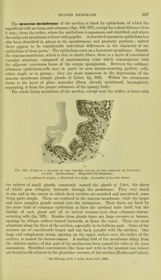
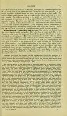
![MUCOUS MEMBRANE ()37 The mucous membrane of the urethra is lined by epithelium, of which the superficial cells are long and columnar (figs. 926,927), except for a short distance (5 to 8 mm.) from the orifice, where the epithelium is squamous and stratified, and where the sub] acent membrane is beset with papillte. A stratified squamous epithelium has also been described in places in the membranous and prostatic portions ; indeed there appear to be considerable individual differences in the character of the epithelium of these parts. ^ The epithelium rests on a basement-membrane. Outside the mucous membrane, which is rich in elastic fibres, there is a layer of convoluted vascular structure, composed of anastomosing veins which communicate with the adjacent cavernous tissue of the corpus spongiosum. Between the ordinary columnar epithelium-cells may in parts be seen mucus-secreting (goblet) cells^ either singly or in groups ; they are most numerous in the depressions of the mucous membrane (simple glands of Littre, fig. 926). Within the submucous tissue is the layer of plain muscular fibres, already mentioned, incompletely separating it from the proper substance of the spongy body. The whole lining membrane of the urethra, except near the orifice, is beset with Fig. 927.—Part of a section op the urethra distal to the orifices of Cowper’s GLANDS. (Lichtenberg.) Magnified 155 diameters. a, b, orifices of crypts; c, blind end of a crypt; the nuclei of its cells shown. the orifices of small glands, commonly named the glands of Littre, the ducts of which pass obliquely forwards through the membrane. They vary much in size and in the extent to which their cavities are ramified and sacculated, some being quite simple. These are confined to the mucous membrane, while the larger and more complex glands extend into the submucosa. Their ducts are lined by the same kind of columnar epithelium as lines the urethral tube itself, but the fundus of each gland and all its lateral recesses have clear columnar mucus- secreting cells (fig. 928). Besides these glands there are large recesses or lacunce, opening by oblique orifices turned forwards, or down the canal. These are most abundant along the floor of the urethra, especially in its bulbous part. Some of the recesses are of considerable length and run back parallel with the urethra. One large and conspicuous recess, opening on the upper surface near the orifice of the urethra, is named the lacuna magna. A median fold of the membrane rising from the inferior surface of this part of the urethra has been named^the valve of the fossa navicularis. Stratified concrements like those met with in the prostate (see below), are found in old subjects in the glandular recesses of the urethra (Robin and Cadiat). 1 See Herzog, Arcb. f. mikr. Anal. Ixiii. 1904,](https://iiif.wellcomecollection.org/image/b21966916_0697.jp2/full/800%2C/0/default.jpg)