The people's common sense medical adviser in plain English, or, Medicine simplified / by R.V. Pierce.
- Ray V. Pierce
- Date:
- 1890
Licence: Public Domain Mark
Credit: The people's common sense medical adviser in plain English, or, Medicine simplified / by R.V. Pierce. Source: Wellcome Collection.
Provider: This material has been provided by the University of Massachusetts Medical School, Lamar Soutter Library, through the Medical Heritage Library. The original may be consulted at the Lamar Soutter Library at the University of Massachusetts Medical School.
30/1034 page 22
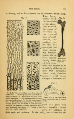
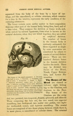
![32 separated from the body of the bone by a layer of car- tilage, and the cancellated, or cellular structure, which remains for a time in the interior, represents the early condition of the ossifying substances. The bones contain more earthy matter in their composition than any other part of the human body, being firm, hard, and of a lime color. They compose the skeleton or frame work, and, when united by natural ligaments, form what is known as the natural skeleton; when they are wired together, they are called an artificial skeleton. ^9' ^^- ' . The number of bones in the human body is variously estimated; for those regarded as single by some anatomists are considered by others to consist of several distinct ])iec'es. There ai*e two hundred distinct bones in the human skeleton besides the teeth. These may be divided into those of the Head, Trunk, Upper Extrem- ities, and Lower Ex- tremities. The Bones of the Head are classed as follows: eight belong- ing to the Cranium, and fourteen to the Face. The bones of the Cranium are the occip- ital, two parietal, two temi^oral, frontal, sphenoid, and ethmoid. Those composing the face are, the two nasal, two superior maxillary, two lachrymal, two malar two palate, two infer- ior turbinated, vomer, and inferior maxillary. The cranial bones are composed of two dense plates, between which there is, in most places, a cancellated or cellular tissue. The external Tho bones of the skull separated. 1. Frontal, only half is seen. 2. Parietal. 3. Occipital, only half is seen. h. Temporal. 5. Nasal. 6. Malar. 7. Superior maxUlary (upper jaw). 8. Lachry- mal. 9. Inferior maxillary (lower jaw). Between 4 and G a part of the sphenoid or wedge-shaped bone, is seen. Another bone assisting to form the slcull, but not here seen, is called tho ethmoid (sieve-like, from being full of holes), and is situ- ated between the sockets of the eyes, forming the roof of the nose.](https://iiif.wellcomecollection.org/image/b21197775_0030.jp2/full/800%2C/0/default.jpg)