A practical treatise on the diseases of the eye / by William Mackenzie ; to which is prefixed an anatomical introduction explanatory of a horizontal section of the human eyeball by Thomas Wharton Jones.
- Date:
- 1840
Licence: Public Domain Mark
Credit: A practical treatise on the diseases of the eye / by William Mackenzie ; to which is prefixed an anatomical introduction explanatory of a horizontal section of the human eyeball by Thomas Wharton Jones. Source: Wellcome Collection.
Provider: This material has been provided by the Royal College of Physicians of Edinburgh. The original may be consulted at the Royal College of Physicians of Edinburgh.
121/978
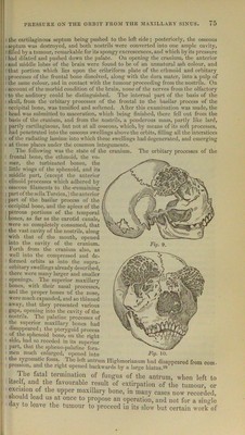
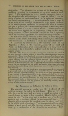
![i 1 : the cartilaginous septum being pushed to the left side; posteriorly, the osseous ! septum was destroyed, and both nostrils were converted into one ample cavity, t filled by a tumour, remarkable for its spongy excrescences, and which by its pressure 1 had dilated and pushed down the palate. On opening the cranium, the anterior ! and middle lobes of the brain were found to be of an unnatural ash colour, and ithat portion which lies upon the cribriform plate of the ethmoid and orbitary j)rocesses of the frontal bone dissolved, along with the dura mater, into a pulp of the same colour, and in contact with the tumour proceeding from the nostrils. On account of the morbid condition of the brain, none of the nerves from the olfactory to the auditory could be distinguished. The internal part of the basis of the skull, from the orbitary processes of the frontal to the basilar process of the occipital bone, was tumified and softened. After this examination was made, the head was submitted to maceration, which being finished, there fell out from the basis of the cranium, and from the nostrils, a ponderous mass, partly like lard, Eartly cartilaginous, but not at all osseous, which, by means of its soft ])rocesses, ad penetrated into the osseous swellings above the orbits, tilting all the interstices of the radiating laminm into which these swellings had degenerated, and emerging at these places under the common integuments. The following was the state of the cranium. The orbitary processes of the frontal bone, the ethmoid, the vo- mer, the turbinated bones, the little wings of the sphenoid, and its middle part, (excej)t the anterior clinoid processes which adhered by osseous filaments to the •remaining part of the sella Turcica,) the anterior part of the basilar process of the occipital bone, and the apices of the petrous portions of the temporal bones, as far as the carotid canals, were so completely consumed, that the vast cavitjr of the nostrils, along with that of the mouth, opened into the cavity of the cranium. Forth from the cranium also, as well into the compressed and de- formed orbits as into the supra- orbitary swellings already described, there were many larger and smaller openings. The superior maxillary bones, with their nasal processes, and the proper bones of the nose, were much expanded, and so thinned away, that they ])resented various gaps, opening into the cavity of the nostrils. The ]ialatine processes of the superior maxillary bones had disappeared; the pterygoid process of the sphenoid bone, on the right side, had so receded in its superior part, that the spheno-palatine fora- men much enlarged, opened into the zygomatic fossa. The left antrum Highmorianum had disappeared from com. pression, and the right opened backwards by a large hiatus.‘-^8 The fatal termination of fungus of the antrum, when left to itselt, and the favourable result of extirpation of the tumour, or excision of the upper maxillary bone, in many cases now recorded, should lead us at once to propose an operation, and not for a single day to leave the tumour to proceed in its slow but certain work of Fig. 9. Fig* 10. 1](https://iiif.wellcomecollection.org/image/b28043467_0121.jp2/full/800%2C/0/default.jpg)