A treatise on the human skeleton (including the joints) / by George Murray Humphry.
- George Murray Humphry
- Date:
- 1858
Licence: Public Domain Mark
Credit: A treatise on the human skeleton (including the joints) / by George Murray Humphry. Source: Wellcome Collection.
Provider: This material has been provided by the Francis A. Countway Library of Medicine, through the Medical Heritage Library. The original may be consulted at the Francis A. Countway Library of Medicine, Harvard Medical School.
643/754 (page 533)
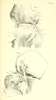
![gastrocnemius; the lower and and foremost (0) gives attachment to the tendon of the jyo-plitcBus, and the middle one [B) gives attachment to the External lateral ligament. This ligament is of cylindrical shape, and 2^ inches long; it is attached, below, to the depression on the head of the fibula, which is just external to the middle of its articular facet, and which is partly surrounded by the prominence for the insertion of the biceps tendon. The points of attachment of the ligament stand out more wide of the joint than do those of the internal lateral ligament; and it is separated fi-om the joint, from the interarticular cartilage, and from the head of the tibia, by an interval containing fat and areolar tissue. The tendon of the ]3oplitceus (0) intervenes between it and the lower edge of the femm, except in the completely flexed state of the joint, when the tendon lies along its own groove. Some of the fibres of the hicejis tendon pass over the lower part of the ligament, in their com'se to the fore part of the fibula and to the adjacent part of the tibia. The tendon is closely united to the ligament by tough fibro- areolar tissue, so as to exert an influence in giving it some tension while the joint is being bent. The external lateral ligament, therefore, differs from tweenthetwo the internal in the following respects. It is of rope- mente ^^^ ^^'^ sliape; is an inch shorter; the point of its attach- ment to the fibula is higher than that of the internal ligament to the tibia; and its point of attachment to the condyle of the femur is a little lower and a little further back^; it is more free from the joint; and has no direct connection either with the head of the tibia or with the interarticular cartilage. Both lateral ligaments lie nearer to the hinder than to iiiost^reia™ed ^^^ ^^rc part of the joint, which alone is sufficient to when joint is xxidkj^ them tifflit in the extended, and relaxed in the bent. ° ' flexed, position of the knee. The relaxation is more marked in the external ligament than in the internal, which per- mits to the external condyle of the tibia the free movement required 1 A pair of compasses will shew this at once, and will prove that the sweep of the outer condyle round the point of attachment of the external lateral ligament is smaller —is part of a smaller circle—than that of the inner condyle round the corresponding point of attachment of the internal lateral ligament.](https://iiif.wellcomecollection.org/image/b21060058_0643.jp2/full/800%2C/0/default.jpg)