Volume 1
The works of Francis Maitland Balfour / edited by M. Foster and Adam Sedgwick.
- Balfour, Francis M. (Francis Maitland), 1851-1882.
- Date:
- 1885
Licence: Public Domain Mark
Credit: The works of Francis Maitland Balfour / edited by M. Foster and Adam Sedgwick. Source: Wellcome Collection.
926/948
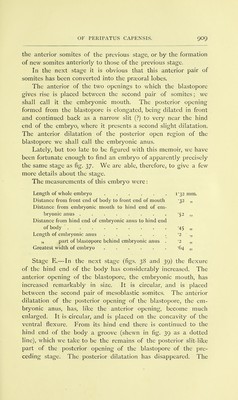
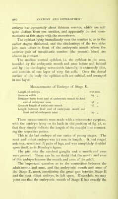
![apex of a well-developed white papilla (PL 47, fig. 4). It is enormously enlarged, and is prolonged forward as a long tubular gland, the structure of which resembles that of the vesicles of the crural glands in the other legs. This gland lies in the lateral compartment of the body cavity, and extends forward to the level of the 9th leg (PL 48, fig. 8, and PL 53, fig. 43). It is described by Professor Balfour as the accessory gland of the male, and is seen in section lying immediately dorsal to the nerve-cord in fig. 20, a^:] PART III., The Development of Peripatus capensis. [The remarkable discoveries about the early development of Peripatus, which Balfour made in June last, shortly before starting for Switzerland, have already been the subject of a short communication to the Royal Society [Proc. Roy. Soc. No. 222, 1882). They relate (i) to the blastopore, (2) to the origin of the mesoblast. Balfour left no manuscript account or notes of his discovery in connection with the drawings which he prepared in order to illustrate it, but he spoke about it to Professor Ray Lankester and also to us, and he further gave a short account of the matter in a private letter to Professor Kleinenberg. In this letter, which by the courtesy of Professor Kleinenberg we have been permitted to see, he describes the blastopore as an elongated slit-like structure extending along nearly the whole ventral surface ; and further states, as the result of his examin- ation of the few and ill-preserved embryos in his possession, that the mesoblast appears to originate as paired outgrowths from the lips of the blastopore. The drawings left by Balfour in connection with the dis- coveries are four in number: one of the entire embryo, shewing the slit-like blastopore and the mesoblastic somites, the other three depicting the transverse sections of the same embryo.](https://iiif.wellcomecollection.org/image/b20417342_001_0930.jp2/full/800%2C/0/default.jpg)