The central course of the nervus octavus and its influence on motility / by C. Winkler.
- Cornelis Winkler
- Date:
- 1907
Licence: In copyright
Credit: The central course of the nervus octavus and its influence on motility / by C. Winkler. Source: Wellcome Collection.
Provider: This material has been provided by King’s College London. The original may be consulted at King’s College London.
184/256 page 180
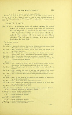
![pulv. th. opt. = pulvinar thalami o])tici. c. p. — commissura posterior. g. hab. — ganglion habenulae. g. interp. — ganglion interpedunculare. N. R. nucleas ruber. Dec. Br. conj. = Decussatio braccli. conjunct, penduncul. cereb. superioris. Plate Xll. Fig. 17 A, B, and c. Frontal sections throngh the medulla oblon- gata of a not yet born elder foetus of a rabbit, (a Weigert-Pat. preparation) showing how far niyelinisation of fibres has taken place. A is the most distal, C the most proximal section. The abbreviations found here are the same as in pre- ceeding drawings. In fig. 17 a. The non medullated layer /? is found between the two medullated la3'ers of the systema dorsale (stria medullaris) nervi octavi. In lig. 17 b. The non medullated layer (stratum c corp. tr.) is found between the medullated layers of the systema ventrale nervi octavi. In fig. 17 c. The medullated triangular iield of the fasciculus spino-cerebellaris ascendens, is maling up to take its dorsal way in the lateral fillet. Plate XIII. Fig. 18 A—-p. A series of sagittal sections through the central system of a not yet born elder foetus of a rabbit (a Weigert-Pal preparation) showing the niyelinisation of the fibres at the time of birth. The abbreviations found here are the same as in the preceeding drawings. Eig. 18 a is a sagittal section just touching the lateral surface of the medulla. Most proximally the nervus trigeminus enters, distally at first the proximal octavus-root is seen, afterwards the distal octavus-root enters. Between them is found the fasc. intermedins radicum. Fig. 18 b is a dito, somewhat more medially. The corpus trapezoides begins its exfoliation from the nucleus ventralis. The fasciculus intermedins is seen. Fig. 18 c is a sagittal section, at the level where the dorsal spino-cerebellar tract (Flechsig's KleinHirn-SeitenstrangBundel) enters into tlie cerebellum. It is medullated, and crossed by medullated fibres of the ventral root going to the stratum latero-dorsale. Fig. 18 D is a sagittal section, at the most medial region of the area ovalis, or at the most lateral region of the corpus juxta-restiforme. The medullated Held's intermediary system is touched as a bundle of longitudinal fibres, passing from the strat. latero-dorsale into the corp. trapezoides. Fig. 18 E is a sagittal section, at the level of the facial nucleus. Held's system now is a field of queer-sectioned small fibres. The section demonstrates the beginning of the tractus Deitehs descendens. Fig. 18 p is a sagittal section through the lateral end ofthegenuNervifacialis.lt demonstrates the curvation in longitudinal direction of the tractus Deiters descendens. Platf, XIV and Plate XV.](https://iiif.wellcomecollection.org/image/b21295712_0184.jp2/full/800%2C/0/default.jpg)




 No text description is available for this image
 No text description is available for this image
 No text description is available for this image