The central course of the nervus octavus and its influence on motility / by C. Winkler.
- Cornelis Winkler
- Date:
- 1907
Licence: In copyright
Credit: The central course of the nervus octavus and its influence on motility / by C. Winkler. Source: Wellcome Collection.
Provider: This material has been provided by King’s College London. The original may be consulted at King’s College London.
188/256 page 184
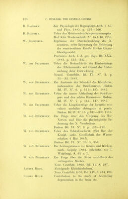
![Fig. 2) E. The section touches at the right side the lesion. The peilunculus cerebelli inferior (its oval area) being cleft, this oval area is degenerating towards the cerebellum. Transverse fibres unite the two ascending Deiters tracts. The medial bundle of the fillet touches the lateral bundle at the dorsal top of the distal end of the nucleus ventralis lemnisci. The systema ven- trale nervi octavi is degenerated. Plate XX. Fig. 25 F. The section touches the lesion in x on both sides. Degenerated are I'y the fasciculus longitudinalis posterior 2'y the tractus Deitees ascendens. S^y MoNAKOw's decussation of transverse fibres going to the dorsal layer of the oliva superior (medial bundle of the fillet), i^y The systema ventrale nervi octavi. Fig. 2.5 G. The section touching still in x the double-sided lesion falls through the exit of the VIF'^ nerve. MoNAKOw's transverse fibres towards the dorsal top of the nucleus olivaris superior are totally degenerate. Fig. 2.5 H. The section touches the tubercalum acusticum and the nucleus ventralis. Degeneration is found in. I'y. The systema dorsale (Monakow's stria medul- laris). 2'y. Held's intermedial system. 3'y. The tractus Deitebs descen- dens. 4'y. The systema ventrale. Fig. 25 I. The section touches the nucleus facialis and the distal end of the tuber- culum acusticum. On both sides is found degeneration in lb' the tractus Deiteks desceudens. 2iy. The fasc. long. post, and the fasciculus praedor- salis, S'y in the fasc. rubro-spinalis 4'y in the nucl. facialis, 5^'in the fibr. transversae dorsalis and 6'y in the radix desceudens N. VHl. Fig. 25 K. The section falls through the Xllt'^ nucleus, demonstrating the degene- ration in the tracts descending towards the cord. Fig. 25 L. The section through the distal end of the med. oblongata. The position of the tractus Deiters desceudens, the fasciculus praedorsalis and the rubrospinal tract. Plate XXI. Fig. 25 M N and o. Sections through ditterent levels of tJie cervical cord(C]^, Cg , Cj) to demonstrate the position of the f. praedorsalis, the descending Deiters tract, and the rubro-spinal tract. Plate XXI and Plate XXII. Fig. 28 A—F and tig. 12. Series of sections through the medulla oblongata of the rabbit, six months after the ablation of the left tuberculum acusticum made in the young born animal. The lesion is found in lig. 28 in a—d. The nucleus ventralis is ablated, but the octavus-roots have but a slight atrophy. The series is drawn to demonstrate the atrophy of the systems in the fillet. (Weigert-Pal preparation). Plate XXL Fig 12. A part of the section in fig. 28 b, limited by a circle, is drawn by an enlargement of '^/i- The nucleus ventralis and the larger part of the corpus trapezoides being lost, it is seen, that fibres of the octavus-root (the dorsal root) pass immediately into the corpus trapezoides. Fig. 28 A. The .section tlirough the distal end of the tub. acusticum.](https://iiif.wellcomecollection.org/image/b21295712_0188.jp2/full/800%2C/0/default.jpg)

 No text description is available for this image
 No text description is available for this image
 No text description is available for this image